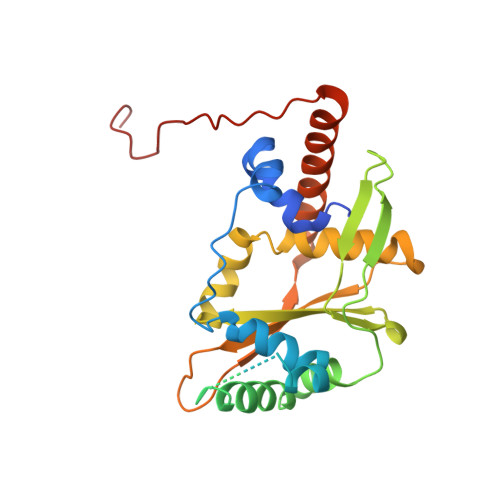
3H4R

Crystal structure of E. coli RecE exonuclease


Experimental Data Snapshot

  • Method: X-RAY DIFFRACTION
  • Resolution: 2.80 Å
  • R-Value Free: 0.313 
  • R-Value Work: 0.290 
  • R-Value Observed: 0.290 

wwPDB Validation   3D Report Full Report


This is version 1.4 of the entry. See complete history


Literature

Crystal structure of E. coli RecE protein reveals a toroidal tetramer for processing double-stranded DNA breaks.

Zhang, J.Xing, X.Herr, A.B.Bell, C.E.

(2009) Structure 17: 690-702

  • DOI: https://doi.org/10.1016/j.str.2009.03.008
  • Primary Citation of Related Structures:  
    3H4R

  • PubMed Abstract: 
  • Escherichia coli RecE protein is part of the classical RecET recombination system that has recently been used in powerful new methods for genetic engineering. RecE binds to free double-stranded DNA (dsDNA) ends and processively digests the 5'-ended strand to form 5'-mononucleotides and a 3'-overhang that is a substrate for single strand annealing promoted by RecT ...

    Escherichia coli RecE protein is part of the classical RecET recombination system that has recently been used in powerful new methods for genetic engineering. RecE binds to free double-stranded DNA (dsDNA) ends and processively digests the 5'-ended strand to form 5'-mononucleotides and a 3'-overhang that is a substrate for single strand annealing promoted by RecT. Here, we report the crystal structure of the C-terminal nuclease domain of RecE at 2.8 A resolution. RecE forms a toroidal tetramer with a central tapered channel that is wide enough to bind dsDNA at one end, but is partially plugged at the other end by the C-terminal segment of the protein. Four narrow tunnels, one within each subunit of the tetramer, lead from the central channel to the four active sites, which lie about 15 A from the channel. The structure, combined with mutational studies, suggests a mechanism in which dsDNA enters through the open end of the central channel, the 5'-ended strand passes through a tunnel to access one of the four active sites, and the 3'-ended strand passes through the plugged end of the channel at the back of the tetramer.


    Organizational Affiliation

    Department of Molecular and Cellular Biochemistry, The Ohio State University College of Medicine, Columbus, OH 43210, USA.


Macromolecules
Find similar proteins by:  (by identity cutoff)  |  3D Structure
Entity ID: 1
MoleculeChainsSequence LengthOrganismDetailsImage
Exodeoxyribonuclease 8265Escherichia coli K-12Mutation(s): 1 
Gene Names: b1350JW1344recE
EC: 3.1.11
UniProt
Find proteins for P15032 (Escherichia coli (strain K12))
Explore P15032 
Go to UniProtKB:  P15032
Entity Groups  
Sequence Clusters30% Identity50% Identity70% Identity90% Identity95% Identity100% Identity
UniProt GroupP15032
Protein Feature View
Expand
  • Reference Sequence
Experimental Data & Validation

Experimental Data

  • Method: X-RAY DIFFRACTION
  • Resolution: 2.80 Å
  • R-Value Free: 0.313 
  • R-Value Work: 0.290 
  • R-Value Observed: 0.290 
  • Space Group: P 4 21 2
Unit Cell:
Length ( Å )Angle ( ˚ )
a = 123.2α = 90
b = 123.2β = 90
c = 67.26γ = 90
Software Package:
Software NamePurpose
MAR345data collection
PHENIXmodel building
CNSrefinement
MOSFLMdata reduction
SCALAdata scaling
PHENIXphasing

Structure Validation

View Full Validation Report


Entry History 

Deposition Data

Revision History  (Full details and data files)

  • Version 1.0: 2009-05-26
    Type: Initial release
  • Version 1.1: 2011-07-13
    Changes: Source and taxonomy, Version format compliance
  • Version 1.2: 2011-12-07
    Changes: Database references
  • Version 1.3: 2017-11-01
    Changes: Refinement description
  • Version 1.4: 2021-10-13
    Changes: Database references