


Crystal Structure of Serine Acetyltransferase CysE from Yersinia pestis
Kim, Y., Zhou, M., Peterson, S., Anderson, W.F., Joachimiak, A.To be published.
Experimental Data Snapshot
Starting Model: experimental
View more details
wwPDB Validation 3D Report Full Report

Entity ID: 1 | |||||
|---|---|---|---|---|---|
| Molecule | Chains | Sequence Length | Organism | Details | Image |
| Serine acetyltransferase | 276 | Yersinia pestis | Mutation(s): 0 Gene Names: cysE, YPO0070 EC: 2.3.1.30 | 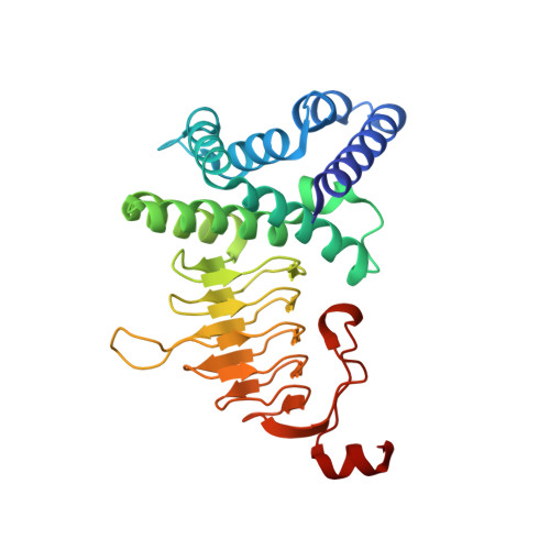 | |
UniProt | |||||
Entity Groups | |||||
| Sequence Clusters | 30% Identity50% Identity70% Identity90% Identity95% Identity100% Identity | ||||
| UniProt Group | A0A2U2H3H7 | ||||
Sequence AnnotationsExpand | |||||
Reference Sequence | |||||
| Ligands 6 Unique | |||||
|---|---|---|---|---|---|
| ID | Chains | Name / Formula / InChI Key | 2D Diagram | 3D Interactions | |
| PG5 Download:Ideal Coordinates CCD File | S [auth C] | 1-METHOXY-2-[2-(2-METHOXY-ETHOXY]-ETHANE C8 H18 O4 YFNKIDBQEZZDLK-UHFFFAOYSA-N |  | ||
| CYS Download:Ideal Coordinates CCD File | AA [auth G] DA [auth H] EA [auth I] IA [auth J] MA [auth L] | CYSTEINE C3 H7 N O2 S XUJNEKJLAYXESH-REOHCLBHSA-N |  | ||
| PEG Download:Ideal Coordinates CCD File | LA [auth L] | DI(HYDROXYETHYL)ETHER C4 H10 O3 MTHSVFCYNBDYFN-UHFFFAOYSA-N |  | ||
| SO4 Download:Ideal Coordinates CCD File | BA [auth H], JA [auth K], KA [auth L], M [auth A], Y [auth F] | SULFATE ION O4 S QAOWNCQODCNURD-UHFFFAOYSA-L |  | ||
| GOL Download:Ideal Coordinates CCD File | CA [auth H], FA [auth J], Q [auth B], W [auth E], Z [auth F] | GLYCEROL C3 H8 O3 PEDCQBHIVMGVHV-UHFFFAOYSA-N |  | ||
| ACY Download:Ideal Coordinates CCD File | GA [auth J], HA [auth J], N [auth A], T [auth C] | ACETIC ACID C2 H4 O2 QTBSBXVTEAMEQO-UHFFFAOYSA-N |  | ||
| Length ( Å ) | Angle ( ˚ ) |
|---|---|
| a = 68.472 | α = 97.07 |
| b = 81.228 | β = 95.32 |
| c = 139.406 | γ = 94.72 |
| Software Name | Purpose |
|---|---|
| SBC-Collect | data collection |
| HKL-3000 | data collection |
| MOLREP | phasing |
| HKL-3000 | phasing |
| PHENIX | refinement |
| HKL-3000 | data reduction |
| HKL-3000 | data scaling |


