3GQI

Crystal Structure of activated receptor tyrosine kinase in complex with substrates


Experimental Data Snapshot

  • Method: X-RAY DIFFRACTION
  • Resolution: 2.50 Å
  • R-Value Free: 0.289 
  • R-Value Work: 0.249 

wwPDB Validation   3D Report Full Report


Ligand Structure Quality Assessment 


This is version 1.4 of the entry. See complete history


Literature

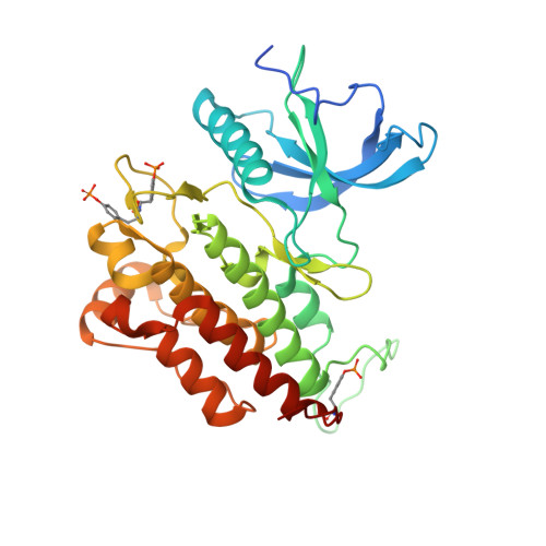
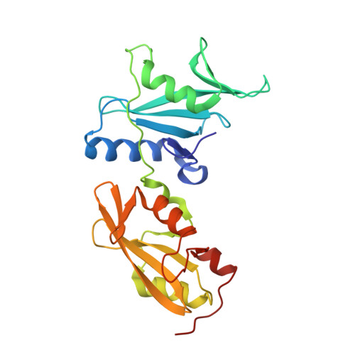
The selectivity of receptor tyrosine kinase signaling is controlled by a secondary SH2 domain binding site.

Bae, J.H.Lew, E.D.Yuzawa, S.Tome, F.Lax, I.Schlessinger, J.

(2009) Cell 138: 514-524

  • DOI: https://doi.org/10.1016/j.cell.2009.05.028
  • Primary Citation of Related Structures:  
    3GQI, 3GQL

  • PubMed Abstract: 
  • SH2 domain-mediated interactions represent a crucial step in transmembrane signaling by receptor tyrosine kinases. SH2 domains recognize phosphotyrosine (pY) in the context of particular sequence motifs in receptor phosphorylation sites. However, the modest binding affinity of SH2 domains to pY containing peptides may not account for and likely represents an oversimplified mechanism for regulation of selectivity of signaling pathways in living cells ...

    SH2 domain-mediated interactions represent a crucial step in transmembrane signaling by receptor tyrosine kinases. SH2 domains recognize phosphotyrosine (pY) in the context of particular sequence motifs in receptor phosphorylation sites. However, the modest binding affinity of SH2 domains to pY containing peptides may not account for and likely represents an oversimplified mechanism for regulation of selectivity of signaling pathways in living cells. Here we describe the crystal structure of the activated tyrosine kinase domain of FGFR1 in complex with a phospholipase Cgamma fragment. The structural and biochemical data and experiments with cultured cells show that the selectivity of phospholipase Cgamma binding and signaling via activated FGFR1 are determined by interactions between a secondary binding site on an SH2 domain and a region in FGFR1 kinase domain in a phosphorylation independent manner. These experiments reveal a mechanism for how SH2 domain selectivity is regulated in vivo to mediate a specific cellular process.


    Organizational Affiliation

    Department of Pharmacology, Yale University School of Medicine, 333 Cedar Street, New Haven, CT 06520, USA.


Macromolecules
Find similar proteins by:  (by identity cutoff)  |  3D Structure
Entity ID: 1
MoleculeChainsSequence LengthOrganismDetailsImage
Basic fibroblast growth factor receptor 1A326Homo sapiensMutation(s): 4 
Gene Names: FGFBRFGFR1FLGFLT2BFGFRCEKHBGFR
EC: 2.7.10.1
UniProt & NIH Common Fund Data Resources
Find proteins for P11362 (Homo sapiens)
Explore P11362 
Go to UniProtKB:  P11362
PHAROS:  P11362
Entity Groups  
Sequence Clusters30% Identity50% Identity70% Identity90% Identity95% Identity100% Identity
UniProt GroupP11362
Protein Feature View
Expand
  • Reference Sequence
Find similar proteins by:  (by identity cutoff)  |  3D Structure
Entity ID: 2
MoleculeChainsSequence LengthOrganismDetailsImage
Phospholipase C-gamma-1B226Rattus norvegicusMutation(s): 0 
Gene Names: Plcg1
EC: 3.1.4.11
UniProt
Find proteins for P10686 (Rattus norvegicus)
Explore P10686 
Go to UniProtKB:  P10686
Entity Groups  
Sequence Clusters30% Identity50% Identity70% Identity90% Identity95% Identity100% Identity
UniProt GroupP10686
Protein Feature View
Expand
  • Reference Sequence
Small Molecules
Ligands 3 Unique
IDChainsName / Formula / InChI Key2D Diagram3D Interactions
DVT
Query on DVT

Download Ideal Coordinates CCD File 
C [auth A]DECAVANADATE
O28 V10
WOYKCMUEHQAMTC-UHFFFAOYSA-N
 Ligand Interaction
ACP
Query on ACP

Download Ideal Coordinates CCD File 
D [auth A]PHOSPHOMETHYLPHOSPHONIC ACID ADENYLATE ESTER
C11 H18 N5 O12 P3
UFZTZBNSLXELAL-IOSLPCCCSA-N
 Ligand Interaction
MG
Query on MG

Download Ideal Coordinates CCD File 
E [auth A]MAGNESIUM ION
Mg
JLVVSXFLKOJNIY-UHFFFAOYSA-N
 Ligand Interaction
Modified Residues  1 Unique
IDChainsTypeFormula2D DiagramParent
PTR
Query on PTR
A L-PEPTIDE LINKINGC9 H12 N O6 PTYR
Experimental Data & Validation

Experimental Data

  • Method: X-RAY DIFFRACTION
  • Resolution: 2.50 Å
  • R-Value Free: 0.289 
  • R-Value Work: 0.249 
  • Space Group: C 1 2 1
Unit Cell:
Length ( Å )Angle ( ˚ )
a = 133.935α = 90
b = 64.596β = 96.5
c = 94.209γ = 90
Software Package:
Software NamePurpose
DENZOdata reduction
SCALEPACKdata scaling
PHASERphasing
CNSrefinement
PDB_EXTRACTdata extraction
HKL-2000data collection
HKL-2000data reduction
HKL-2000data scaling

Structure Validation

View Full Validation Report


Ligand Structure Quality Assessment 


Entry History 

Deposition Data

Revision History  (Full details and data files)

  • Version 1.0: 2009-08-18
    Type: Initial release
  • Version 1.1: 2011-07-13
    Changes: Version format compliance
  • Version 1.2: 2012-03-21
    Changes: Database references
  • Version 1.3: 2017-11-01
    Changes: Refinement description
  • Version 1.4: 2021-10-20
    Changes: Database references, Derived calculations