3GQB

Crystal Structure of the A3B3 complex from V-ATPase


Experimental Data Snapshot

  • Method: X-RAY DIFFRACTION
  • Resolution: 2.80 Å
  • R-Value Free: 0.281 
  • R-Value Work: 0.250 
  • R-Value Observed: 0.251 

wwPDB Validation   3D Report Full Report


This is version 1.3 of the entry. See complete history


Literature

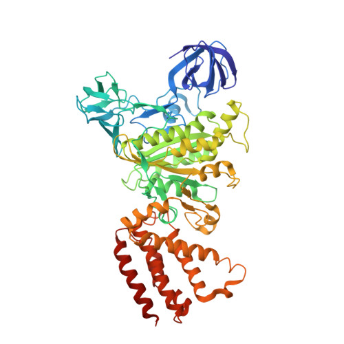
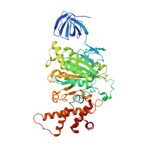
Crystal structure of A(3)B(3) complex of V-ATPase from Thermus thermophilus.

Maher, M.J.Akimoto, S.Iwata, M.Nagata, K.Hori, Y.Yoshida, M.Yokoyama, S.Iwata, S.Yokoyama, K.

(2009) EMBO J 28: 3771-3779

  • DOI: https://doi.org/10.1038/emboj.2009.310
  • Primary Citation of Related Structures:  
    3GQB

  • PubMed Abstract: 

    Vacuolar-type ATPases (V-ATPases) exist in various cellular membranes of many organisms to regulate physiological processes by controlling the acidic environment. Here, we have determined the crystal structure of the A(3)B(3) subcomplex of V-ATPase at 2.8 A resolution. The overall construction of the A(3)B(3) subcomplex is significantly different from that of the alpha(3)beta(3) sub-domain in F(o)F(1)-ATP synthase, because of the presence of a protruding 'bulge' domain feature in the catalytic A subunits. The A(3)B(3) subcomplex structure provides the first molecular insight at the catalytic and non-catalytic interfaces, which was not possible in the structures of the separate subunits alone. Specifically, in the non-catalytic interface, the B subunit seems to be incapable of binding ATP, which is a marked difference from the situation indicated by the structure of the F(o)F(1)-ATP synthase. In the catalytic interface, our mutational analysis, on the basis of the A(3)B(3) structure, has highlighted the presence of a cluster composed of key hydrophobic residues, which are essential for ATP hydrolysis by V-ATPases.


  • Organizational Affiliation

    Division of Molecular Biosciences, Imperial College London, South Kensington Campus, London, UK.


Macromolecules
Find similar proteins by:  (by identity cutoff)  |  3D Structure
Entity ID: 1
MoleculeChains Sequence LengthOrganismDetailsImage
V-type ATP synthase alpha chain
A, C
578Thermus thermophilus HB8Mutation(s): 4 
Gene Names: atpATTHA1273
EC: 3.6.3.14
UniProt
Find proteins for Q56403 (Thermus thermophilus (strain ATCC 27634 / DSM 579 / HB8))
Explore Q56403 
Go to UniProtKB:  Q56403
Entity Groups  
Sequence Clusters30% Identity50% Identity70% Identity90% Identity95% Identity100% Identity
UniProt GroupQ56403
Sequence Annotations
Expand
  • Reference Sequence
Find similar proteins by:  (by identity cutoff)  |  3D Structure
Entity ID: 2
MoleculeChains Sequence LengthOrganismDetailsImage
V-type ATP synthase beta chain
B, D
464Thermus thermophilus HB8Mutation(s): 1 
Gene Names: atpBTTHA1272
UniProt
Find proteins for Q56404 (Thermus thermophilus (strain ATCC 27634 / DSM 579 / HB8))
Explore Q56404 
Go to UniProtKB:  Q56404
Entity Groups  
Sequence Clusters30% Identity50% Identity70% Identity90% Identity95% Identity100% Identity
UniProt GroupQ56404
Sequence Annotations
Expand
  • Reference Sequence
Experimental Data & Validation

Experimental Data

  • Method: X-RAY DIFFRACTION
  • Resolution: 2.80 Å
  • R-Value Free: 0.281 
  • R-Value Work: 0.250 
  • R-Value Observed: 0.251 
  • Space Group: P 3 2 1
Unit Cell:
Length ( Å )Angle ( ˚ )
a = 199.368α = 90
b = 199.368β = 90
c = 179.005γ = 120
Software Package:
Software NamePurpose
HKL-2000data collection
PHASERphasing
REFMACrefinement
HKL-2000data reduction
SCALEPACKdata scaling

Structure Validation

View Full Validation Report


Entry History 

Deposition Data

Revision History  (Full details and data files)

  • Version 1.0: 2009-11-24
    Type: Initial release
  • Version 1.1: 2011-07-13
    Changes: Advisory, Version format compliance
  • Version 1.2: 2021-10-20
    Changes: Database references
  • Version 1.3: 2024-02-21
    Changes: Data collection