3GID | pdb_00003gid

The biotin carboxylase (BC) domain of human Acetyl-CoA Carboxylase 2 (ACC2) in complex with Soraphen A


Experimental Data Snapshot

  • Method: X-RAY DIFFRACTION
  • Resolution: 2.30 Å
  • R-Value Free: 
    0.242 (Depositor), 0.238 (DCC) 
  • R-Value Work: 
    0.200 (Depositor), 0.196 (DCC) 
  • R-Value Observed: 
    0.202 (Depositor) 

wwPDB Validation 3D Report Full Report

Validation slider image for 3GID

Ligand Structure Quality Assessment 


This is version 1.3 of the entry. See complete history

Literature

Synthesis and characterization of a BODIPY-labeled derivative of Soraphen A that binds to acetyl-CoA carboxylase.

Raymer, B.Kavana, M.Price, A.Wang, B.Corcoran, L.Kulathila, R.Groarke, J.Mann, T.

(2009) Bioorg Med Chem Lett 19: 2804-2807

  • DOI: https://doi.org/10.1016/j.bmcl.2009.03.107
  • Primary Citation Related Structures: 
    3GID, 3GLK

  • PubMed Abstract: 

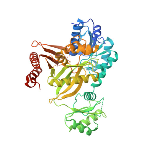
    BODIPY-labeled Soraphen A derivative 4 was synthesized and characterized as an acetyl-CoA carboxylase (ACC) binder. Biophysical measurements indicate that the molecule binds in the biotin carboxylase domain where Soraphen A has been shown to bind. The fluorescent label of the BODIPY can be used to biophysically identify a compound that binds to the Soraphen A site of the biotin carboxylase domain versus the carboxytransferase domain of ACC.


  • Organizational Affiliation
    • Novartis Institutes for BioMedical Research, Cambridge, MA 02139, United States. brian.raymer@novartis.com

Macromolecule Content 

  • Total Structure Weight: 121.23 kDa 
  • Atom Count: 7,413 
  • Modeled Residue Count: 917 
  • Deposited Residue Count: 1,080 
  • Unique protein chains: 1

Macromolecules

Find similar proteins by:|  3D Structure
Entity ID: 1
MoleculeChains  Sequence LengthOrganismDetailsImage
Acetyl-CoA carboxylase 2
A, B
540Homo sapiensMutation(s): 0 
Gene Names: ACACBACC2ACCB
EC: 6.4.1.2 (PDB Primary Data), 6.3.4.14 (PDB Primary Data)
UniProt & NIH Common Fund Data Resources
Find proteins for O00763 (Homo sapiens)
Explore O00763 
Go to UniProtKB:  O00763
PHAROS:  O00763
GTEx:  ENSG00000076555 
Entity Groups
Sequence Clusters30% Identity50% Identity70% Identity90% Identity95% Identity100% Identity
UniProt GroupO00763
Sequence Annotations
Expand
Reference Sequence

Experimental Data & Validation

Experimental Data

  • Method: X-RAY DIFFRACTION
  • Resolution: 2.30 Å
  • R-Value Free:  0.242 (Depositor), 0.238 (DCC) 
  • R-Value Work:  0.200 (Depositor), 0.196 (DCC) 
  • R-Value Observed: 0.202 (Depositor) 
Space Group: C 2 2 21
Unit Cell:
Length ( Å )Angle ( ˚ )
a = 138.339α = 90
b = 141β = 90
c = 123.615γ = 90
Software Package:
Software NamePurpose
CrystalCleardata collection
PHASERphasing
CNXrefinement
HKL-2000data reduction
HKL-2000data scaling

Structure Validation

View Full Validation Report


Ligand Structure Quality Assessment 


Entry History 

Deposition Data

Revision History  (Full details and data files)

  • Version 1.0: 2009-03-17
    Type: Initial release
  • Version 1.1: 2011-07-13
    Changes: Version format compliance
  • Version 1.2: 2014-12-10
    Changes: Database references
  • Version 1.3: 2024-02-21
    Changes: Data collection, Database references, Derived calculations