3GB3 | pdb_00003gb3

X-ray structure of genetically encoded photosensitizer KillerRed in native form


Experimental Data Snapshot

  • Method: X-RAY DIFFRACTION
  • Resolution: 1.75 Å
  • R-Value Free: 
    0.244 (Depositor), 0.245 (DCC) 
  • R-Value Work: 
    0.186 (Depositor), 0.187 (DCC) 
  • R-Value Observed: 
    0.189 (Depositor) 

wwPDB Validation 3D Report Full Report

Validation slider image for 3GB3

This is version 3.0 of the entry. See complete history

Literature

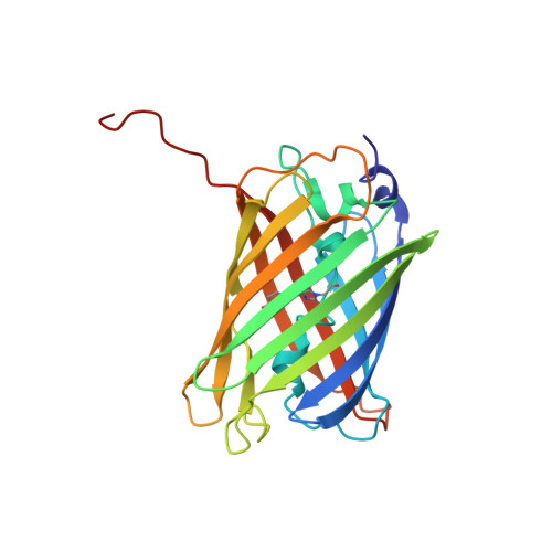
Structural basis for phototoxicity of the genetically encoded photosensitizer KillerRed.

Pletnev, S.Gurskaya, N.G.Pletneva, N.V.Lukyanov, K.A.Chudakov, D.M.Martynov, V.I.Popov, V.O.Kovalchuk, M.V.Wlodawer, A.Dauter, Z.Pletnev, V.

(2009) J Biological Chem 284: 32028-32039

  • DOI: https://doi.org/10.1074/jbc.M109.054973
  • Primary Citation Related Structures: 
    3GB3, 3GL4

  • PubMed Abstract: 

    KillerRed is the only known fluorescent protein that demonstrates notable phototoxicity, exceeding that of the other green and red fluorescent proteins by at least 1,000-fold. KillerRed could serve as an instrument to inactivate target proteins or to kill cell populations in photodynamic therapy. However, the nature of KillerRed phototoxicity has remained unclear, impeding the development of more phototoxic variants. Here we present the results of a high resolution crystallographic study of KillerRed in the active fluorescent and in the photobleached non-fluorescent states. A unique and striking feature of the structure is a water-filled channel reaching the chromophore area from the end cap of the beta-barrel that is probably one of the key structural features responsible for phototoxicity. A study of the structure-function relationship of KillerRed, supported by structure-based, site-directed mutagenesis, has also revealed the key residues most likely responsible for the phototoxic effect. In particular, Glu(68) and Ser(119), located adjacent to the chromophore, have been assigned as the primary trigger of the reaction chain.


  • Organizational Affiliation
    • Synchrotron Radiation Research Section, Macromolecular Crystallography Laboratory, National Cancer Institute/SAIC-Frederick Inc., Frederick, Maryland 21702, USA.

Macromolecule Content 

  • Total Structure Weight: 53.21 kDa 
  • Atom Count: 3,916 
  • Modeled Residue Count: 460 
  • Deposited Residue Count: 470 
  • Unique protein chains: 1

Macromolecules

Find similar proteins by:|  3D Structure
Entity ID: 1
MoleculeChains  Sequence LengthOrganismDetailsImage
KillerRed
A, B
235Anthomedusae sp. DC-2005Mutation(s): 0 
UniProt
Find proteins for Q2TCH5 (Anthomedusae sp. DC-2005)
Explore Q2TCH5 
Go to UniProtKB:  Q2TCH5
Entity Groups
Sequence Clusters30% Identity50% Identity70% Identity90% Identity95% Identity100% Identity
UniProt GroupQ2TCH5
Sequence Annotations
Expand
Reference Sequence

Experimental Data & Validation

Experimental Data

  • Method: X-RAY DIFFRACTION
  • Resolution: 1.75 Å
  • R-Value Free:  0.244 (Depositor), 0.245 (DCC) 
  • R-Value Work:  0.186 (Depositor), 0.187 (DCC) 
  • R-Value Observed: 0.189 (Depositor) 
Space Group: P 21 21 21
Unit Cell:
Length ( Å )Angle ( ˚ )
a = 72.016α = 90
b = 73.682β = 90
c = 78.659γ = 90
Software Package:
Software NamePurpose
DENZOdata reduction
SCALEPACKdata scaling
REFMACrefinement
PDB_EXTRACTdata extraction
SERGUIdata collection
HKL-2000data reduction
HKL-2000data scaling
MOLREPphasing

Structure Validation

View Full Validation Report


Entry History 

Deposition Data

Revision History  (Full details and data files)

  • Version 1.0: 2009-09-08
    Type: Initial release
  • Version 1.1: 2011-07-13
    Changes: Version format compliance
  • Version 1.2: 2017-11-01
    Changes: Refinement description
  • Version 2.0: 2023-11-15
    Changes: Advisory, Atomic model, Data collection, Database references, Derived calculations
  • Version 2.1: 2024-10-09
    Changes: Structure summary
  • Version 3.0: 2026-03-18
    Changes: Polymer sequence