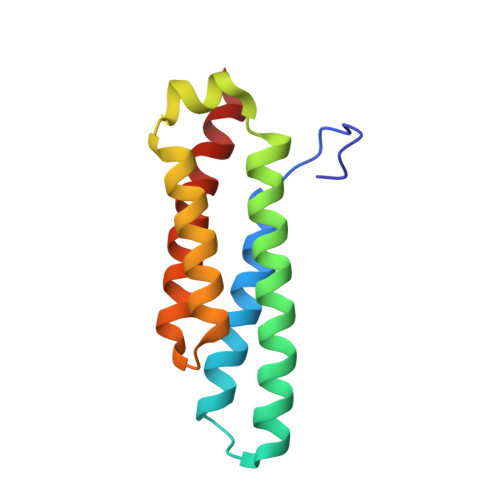
Role of conserved tyrosine residues in NiSOD catalysis: a case of convergent evolution
Herbst, R.W., Guce, A., Bryngelson, P.A., Higgins, K.A., Ryan, K.C., Cabelli, D.E., Garman, S.C., Maroney, M.J.(2009) Biochemistry 48: 3354-3369
- PubMed: 19183068 Search on PubMedSearch on PubMed Central
- DOI: https://doi.org/10.1021/bi802029t
- Primary Citation Related Structures:
3G4X, 3G4Z, 3G50 - PubMed Abstract:
Superoxide dismutases rely on protein structural elements to adjust the redox potential of the metallocenter to an optimum value near 300 mV (vs NHE), to provide a source of protons for catalysis, and to control the access of anions to the active site. These aspects of the catalytic mechanism are examined herein for recombinant preparations of the nickel-dependent SOD (NiSOD) from Streptomyces coelicolor and for a series of mutants that affect a key tyrosine residue, Tyr9 (Y9F-, Y62F-, Y9F/Y62F-, and D3A-NiSOD). Structural aspects of the nickel sites are examined by a combination of EPR and X-ray absorption spectroscopies, and by single-crystal X-ray diffraction at approximately 1.9 A resolution in the case of Y9F- and D3A-NiSODs. The functional effects of the mutations are examined by kinetic studies employing pulse radiolytic generation of O2- and by redox titrations. These studies reveal that although the structure of the nickel center in NiSOD is unique, the ligand environment is designed to optimize the redox potential at 290 mV and results in the oxidation of 50% of the nickel centers in the oxidized hexamer. Kinetic investigations show that all of the mutant proteins have considerable activity. In the case of Y9F-NiSOD, the enzyme exhibits saturation behavior that is not observed in wild-type (WT) NiSOD and suggests that release of peroxide is inhibited. The crystal structure of Y9F-NiSOD reveals an anion binding site that is occupied by either Cl- or Br- and is located close to but not within bonding distance of the nickel center. The structure of D3A-NiSOD reveals that in addition to affecting the interaction between subunits, this mutation repositions Tyr9 and leads to altered chemistry with peroxide. Comparisons with Mn(SOD) and Fe(SOD) reveal that although different strategies for adjusting the redox potential and supply of protons are employed, NiSOD has evolved a similar strategy for controlling the access of anions to the active site.
- Department of Chemistry, UniVersity of Massachusetts, Amherst, Massachusetts 01003, USA.
Organizational Affiliation: