3FX4

Porcine aldehyde reductase in ternary complex with inhibitor


Experimental Data Snapshot

  • Method: X-RAY DIFFRACTION
  • Resolution: 1.99 Å
  • R-Value Free: 0.246 
  • R-Value Work: 0.192 
  • R-Value Observed: 0.194 

wwPDB Validation   3D Report Full Report


Ligand Structure Quality Assessment 


This is version 1.4 of the entry. See complete history


Literature

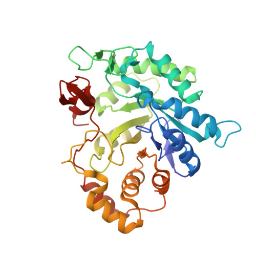
Structure of aldehyde reductase in ternary complex with a 5-arylidene-2,4-thiazolidinedione aldose reductase inhibitor.

Carbone, V.Giglio, M.Chung, R.Huyton, T.Adams, J.Maccari, R.Ottana, R.Hara, A.El-Kabbani, O.

(2010) Eur J Med Chem 45: 1140-1145

  • DOI: https://doi.org/10.1016/j.ejmech.2009.12.019
  • Primary Citation of Related Structures:  
    3FX4

  • PubMed Abstract: 

    The structure of aldehyde reductase (ALR1) in ternary complex with the coenzyme NADPH and [5-(3-carboxymethoxy-4-methoxybenzylidene)-2,4-dioxothiazolidin-3-yl]acetic acid (CMD), a potent inhibitor of aldose reductase (ALR2), was determined at 1.99A resolution. The partially disordered inhibitor formed a tight network of hydrogen bonds with the active site residues (Tyr50 and His113) and coenzyme. Molecular modelling calculations and inhibitory activity measurements of CMD and [5-(3-hydroxy-4-methoxybenzylidene)-2,4-dioxothiazolidin-3-yl]acetic acid (HMD) indicated that pi-stacking interactions with several conserved active site tryptophan residues and hydrogen-bonding interactions with the non-conserved C-terminal residue Leu300 in ALR2 (Pro301 in ALR1) contributed to inhibitor selectivity. In particular for the potent inhibitor CMD, the rotameric state of the conserved residue Trp219 (Trp220 in ALR1) is important in forming a pi-stacking interaction with the inhibitor in ALR2 and contributes to the difference in the binding of the inhibitor to the enzymes.


  • Organizational Affiliation

    Medicinal Chemistry and Drug Action, Monash Institute of Pharmaceutical Sciences, Monash University, 381 Royal Parade, Parkville, Victoria 3052, Australia.


Macromolecules
Find similar proteins by:  (by identity cutoff)  |  3D Structure
Entity ID: 1
MoleculeChains Sequence LengthOrganismDetailsImage
Alcohol dehydrogenase [NADP+]325Sus scrofaMutation(s): 0 
EC: 1.1.1.2
UniProt
Find proteins for P50578 (Sus scrofa)
Explore P50578 
Go to UniProtKB:  P50578
Entity Groups  
Sequence Clusters30% Identity50% Identity70% Identity90% Identity95% Identity100% Identity
UniProt GroupP50578
Sequence Annotations
Expand
  • Reference Sequence
Small Molecules
Binding Affinity Annotations 
IDSourceBinding Affinity
FX4 BindingDB:  3FX4 IC50: 5400 (nM) from 1 assay(s)
Experimental Data & Validation

Experimental Data

  • Method: X-RAY DIFFRACTION
  • Resolution: 1.99 Å
  • R-Value Free: 0.246 
  • R-Value Work: 0.192 
  • R-Value Observed: 0.194 
  • Space Group: P 65 2 2
Unit Cell:
Length ( Å )Angle ( ˚ )
a = 67.35α = 90
b = 67.35β = 90
c = 246.25γ = 120
Software Package:
Software NamePurpose
Blu-Icedata collection
MOLREPphasing
REFMACrefinement
MOSFLMdata reduction
SCALAdata scaling

Structure Validation

View Full Validation Report


Ligand Structure Quality Assessment 


Entry History 

Deposition Data

Revision History  (Full details and data files)

  • Version 1.0: 2009-12-22
    Type: Initial release
  • Version 1.1: 2011-07-13
    Changes: Version format compliance
  • Version 1.2: 2017-11-01
    Changes: Advisory, Refinement description
  • Version 1.3: 2023-10-11
    Changes: Advisory, Data collection, Database references, Derived calculations
  • Version 1.4: 2023-11-01
    Changes: Refinement description