Insights into how nucleotide-binding domains power ABC transport.
Newstead, S., Fowler, P.W., Bilton, P., Carpenter, E.P., Sadler, P.J., Campopiano, D.J., Sansom, M.S., Iwata, S.(2009) Structure 17: 1213-1222
- PubMed: 19748342 Search on PubMedSearch on PubMed Central
- DOI: https://doi.org/10.1016/j.str.2009.07.009
- Primary Citation Related Structures:
3FVQ - PubMed Abstract:
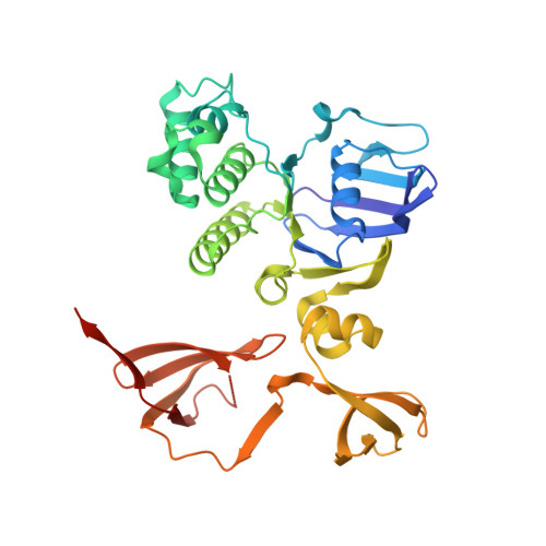
The mechanism by which nucleotide-binding domains (NBDs) of ABC transporters power the transport of substrates across cell membranes is currently unclear. Here we report the crystal structure of an NBD, FbpC, from the Neisseria gonorrhoeae ferric iron uptake transporter with an unusual and substantial domain swap in the C-terminal regulatory domain. This entanglement suggests that FbpC is unable to open to the same extent as the homologous protein MalK. Using molecular dynamics we demonstrate that this is not the case: both NBDs open rapidly once ATP is removed. We conclude from this result that the closed structures of FbpC and MalK have higher free energies than their respective open states. This result has important implications for our understanding of the mechanism of power generation in ABC transporters, because the unwinding of this free energy ensures that the opening of these two NBDs is also powered.
- Division of Molecular Biosciences, Membrane Protein Crystallography Group, Imperial College London, London, UK.
Organizational Affiliation: