3FF5 | pdb_00003ff5

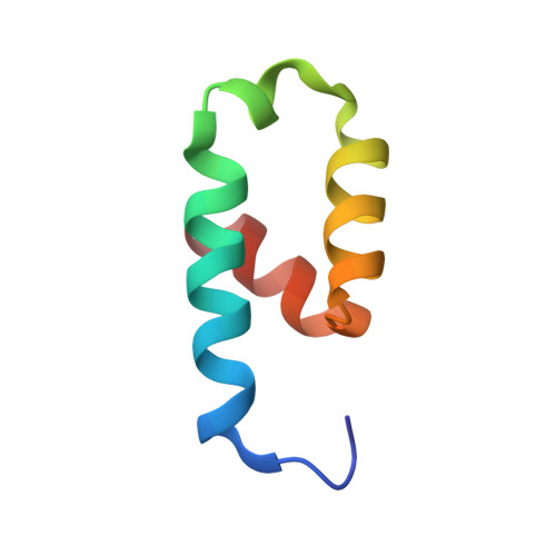
Crystal structure of the conserved N-terminal domain of the peroxisomal matrix-protein-import receptor, Pex14p


Experimental Data Snapshot

  • Method: X-RAY DIFFRACTION
  • Resolution: 1.80 Å
  • R-Value Free: 
    0.211 (Depositor), 0.210 (DCC) 
  • R-Value Work: 
    0.179 (Depositor), 0.180 (DCC) 
  • R-Value Observed: 
    0.181 (Depositor) 

wwPDB Validation   3D Report Full Report


Ligand Structure Quality Assessment 

Created with Raphaël 2.3.0Worse 01 BetterLigand structure goodness of fit to experimental dataBest fitted DPWClick on this verticalbar to view details

This is version 1.3 of the entry. See complete history


Literature

Crystal structure of the conserved N-terminal domain of the peroxisomal matrix protein import receptor, Pex14p

Su, J.R.Takeda, K.Tamura, S.Fujiki, Y.Miki, K.

(2009) Proc Natl Acad Sci U S A 106: 417-421

  • DOI: https://doi.org/10.1073/pnas.0808681106
  • Primary Citation of Related Structures:  
    3FF5

  • PubMed Abstract: 

    Pex14p is a central component of the peroxisomal protein import machinery, in which the conserved N-terminal domain mediates dynamic interactions with other peroxins including Pex5p, Pex13p, and Pex19p. Here, we report the crystal structure of the conserved N-terminal domain of Pex14p with a three-helix bundle. A hydrophobic surface is composed of the conserved residues, of which two phenylalanine residues (Phe-35 and Phe-52) protrude to the solvent. Consequently, two putative binding pockets suitable for recognizing the helical WXXXF/Y motif of Pex5p are formed on the surface by the two phenylalanine residues accompanying with positively charged residues. The structural feature agrees well with our earlier findings where F35A/L36A and F52A/L53A mutants were impaired in the interactions with other peroxins such as Pex5p and Pex13p. Pex14p variants each with Phe-to-Ala mutation at positions 35, 52, and 35/52, respectively, were defective in restoring the impaired peroxisomal protein import in pex14 Chinese hamster ovary mutant ZP161 cells. Moreover, in GST pull-down assays His(6)-Pex5pL bound only to GST-Pex14p(25-70), not to any of GST-Pex14p(25-70)F35A, GST-Pex14p(25-70)F52A, and GST-Pex14p(25-70)F35A/F52A. Endogenous Pex5p was recruited to FLAG-Pex14p on peroxisomes in vivo but barely to FLAG-Pex14pF35A, FLAG-Pex14pF52A, and FLAG-Pex14pF35A/F52A. Collectively, Phe-35 and Phe-52 are essential for the Pex14p functions, including the interaction between Pex14p and Pex5p.


  • Organizational Affiliation

    Department of Chemistry, Graduate School of Science, Kyoto University, Sakyo-ku, Kyoto 606-8502, Japan.


Macromolecules
Find similar proteins by:  (by identity cutoff)  |  3D Structure
Entity ID: 1
MoleculeChains Sequence LengthOrganismDetailsImage
Peroxisomal biogenesis factor 14
A, B
54Rattus norvegicusMutation(s): 0 
Gene Names: Pex14
UniProt
Find proteins for Q642G4 (Rattus norvegicus)
Explore Q642G4 
Go to UniProtKB:  Q642G4
Entity Groups  
Sequence Clusters30% Identity50% Identity70% Identity90% Identity95% Identity100% Identity
UniProt GroupQ642G4
Sequence Annotations
Expand
  • Reference Sequence
Small Molecules
Ligands 1 Unique
IDChains Name / Formula / InChI Key2D Diagram3D Interactions
DPW
Query on DPW

Download Ideal Coordinates CCD File 
C [auth A]decyl 2-trimethylazaniumylethyl phosphate
C15 H34 N O4 P
VVVDGSCGBGBGFN-UHFFFAOYSA-N
Experimental Data & Validation

Experimental Data

  • Method: X-RAY DIFFRACTION
  • Resolution: 1.80 Å
  • R-Value Free:  0.211 (Depositor), 0.210 (DCC) 
  • R-Value Work:  0.179 (Depositor), 0.180 (DCC) 
  • R-Value Observed: 0.181 (Depositor) 
Space Group: I 2 3
Unit Cell:
Length ( Å )Angle ( ˚ )
a = 90.57α = 90
b = 90.57β = 90
c = 90.57γ = 90
Software Package:
Software NamePurpose
SOLVEphasing
REFMACrefinement
HKL-2000data reduction
HKL-2000data scaling

Structure Validation

View Full Validation Report


Ligand Structure Quality Assessment 

Created with Raphaël 2.3.0Worse 01 BetterLigand structure goodness of fit to experimental dataBest fitted DPWClick on this verticalbar to view details

Entry History 

Deposition Data

Revision History  (Full details and data files)

  • Version 1.0: 2008-12-30
    Type: Initial release
  • Version 1.1: 2011-07-13
    Changes: Version format compliance
  • Version 1.2: 2012-05-02
    Changes: Database references
  • Version 1.3: 2023-12-27
    Changes: Data collection, Database references, Derived calculations