3FBF

Crystal structure of the Mimivirus NDK N62L mutant complexed with dTDP


Experimental Data Snapshot

  • Method: X-RAY DIFFRACTION
  • Resolution: 2.60 Å
  • R-Value Free: 0.224 
  • R-Value Work: 0.178 
  • R-Value Observed: 0.181 

wwPDB Validation   3D Report Full Report


Ligand Structure Quality Assessment 


This is version 1.3 of the entry. See complete history


Literature

Dissecting the unique nucleotide specificity of mimivirus nucleoside diphosphate kinase.

Jeudy, S.Lartigue, A.Claverie, J.M.Abergel, C.

(2009) J Virol 83: 7142-7150

  • DOI: https://doi.org/10.1128/JVI.00511-09
  • Primary Citation of Related Structures:  
    2B8P, 2B8Q, 3B6B, 3DDI, 3DKD, 3EE3, 3EIC, 3EJM, 3ELH, 3EM1, 3EMT, 3ENA, 3ETM, 3EVM, 3EVO, 3EVW, 3FBB, 3FBC, 3FBE, 3FBF, 3FC9, 3FCV, 3FCW, 3G2X, 3GP9, 3GPA

  • PubMed Abstract: 

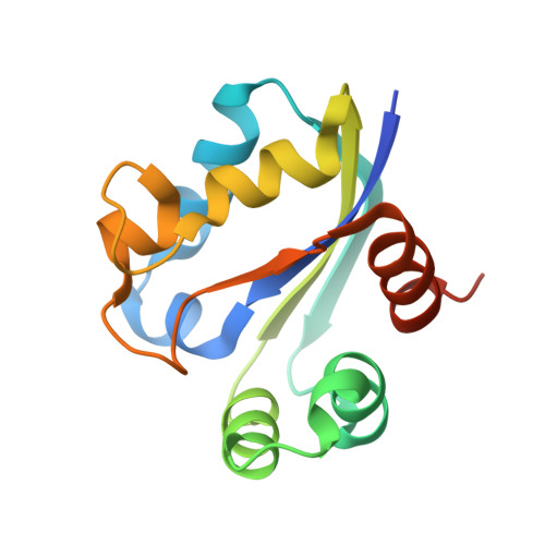
    The analysis of the Acanthamoeba polyphaga mimivirus genome revealed the first virus-encoded nucleoside diphosphate kinase (NDK), an enzyme that is central to the synthesis of RNA and DNA, ubiquitous in cellular organisms, and well conserved among the three domains of life. In contrast with the broad specificity of cellular NDKs for all types of ribo- and deoxyribonucleotides, the mimivirus enzyme exhibits a strongly preferential affinity for deoxypyrimidines. In order to elucidate the molecular basis of this unique substrate specificity, we determined the three-dimensional (3D) structure of the Acanthamoeba polyphaga mimivirus NDK alone and in complex with various nucleotides. As predicted from a sequence comparison with cellular NDKs, the 3D structure of the mimivirus enzyme exhibits a shorter Kpn loop, previously recognized as a main feature of the NDK active site. The structure of the viral enzyme in complex with various nucleotides also pinpointed two residue changes, both located near the active site and specific to the viral NDK, which could explain its stronger affinity for deoxynucleotides and pyrimidine nucleotides. The role of these residues was explored by building a set of viral NDK variants, assaying their enzymatic activities, and determining their 3D structures in complex with various nucleotides. A total of 26 crystallographic structures were determined at resolutions ranging from 2.8 A to 1.5 A. Our results suggest that the mimivirus enzyme progressively evolved from an ancestral NDK under the constraints of optimizing its efficiency for the replication of an AT-rich (73%) viral genome in a thymidine-limited host environment.


  • Organizational Affiliation

    Structural and Genomic Information Laboratory, CNRS-UPR2589, IFR88, Marseille Cedex 9, France.


Macromolecules
Find similar proteins by:  (by identity cutoff)  |  3D Structure
Entity ID: 1
MoleculeChains Sequence LengthOrganismDetailsImage
Nucleoside diphosphate kinase
A, B, C, D, E
A, B, C, D, E, F
142Acanthamoeba polyphaga mimivirusMutation(s): 1 
Gene Names: MIMI_R418NDK
EC: 2.7.4.6
UniProt
Find proteins for Q5UQL3 (Acanthamoeba polyphaga mimivirus)
Explore Q5UQL3 
Go to UniProtKB:  Q5UQL3
Entity Groups  
Sequence Clusters30% Identity50% Identity70% Identity90% Identity95% Identity100% Identity
UniProt GroupQ5UQL3
Sequence Annotations
Expand
  • Reference Sequence
Small Molecules
Ligands 2 Unique
IDChains Name / Formula / InChI Key2D Diagram3D Interactions
TYD
Query on TYD

Download Ideal Coordinates CCD File 
H [auth A]
J [auth B]
L [auth C]
N [auth D]
P [auth E]
H [auth A],
J [auth B],
L [auth C],
N [auth D],
P [auth E],
R [auth F]
THYMIDINE-5'-DIPHOSPHATE
C10 H16 N2 O11 P2
UJLXYODCHAELLY-XLPZGREQSA-N
MG
Query on MG

Download Ideal Coordinates CCD File 
G [auth A]
I [auth B]
K [auth C]
M [auth D]
O [auth E]
G [auth A],
I [auth B],
K [auth C],
M [auth D],
O [auth E],
Q [auth F]
MAGNESIUM ION
Mg
JLVVSXFLKOJNIY-UHFFFAOYSA-N
Experimental Data & Validation

Experimental Data

  • Method: X-RAY DIFFRACTION
  • Resolution: 2.60 Å
  • R-Value Free: 0.224 
  • R-Value Work: 0.178 
  • R-Value Observed: 0.181 
  • Space Group: C 2 2 21
Unit Cell:
Length ( Å )Angle ( ˚ )
a = 80.241α = 90
b = 152.68β = 90
c = 185.248γ = 90
Software Package:
Software NamePurpose
ADSCdata collection
AMoREphasing
PHENIXrefinement
MOSFLMdata reduction
SCALAdata scaling

Structure Validation

View Full Validation Report


Ligand Structure Quality Assessment 


Entry History 

Deposition Data

Revision History  (Full details and data files)

  • Version 1.0: 2009-08-11
    Type: Initial release
  • Version 1.1: 2011-07-13
    Changes: Version format compliance
  • Version 1.2: 2021-10-20
    Changes: Database references, Derived calculations
  • Version 1.3: 2023-09-06
    Changes: Data collection, Refinement description