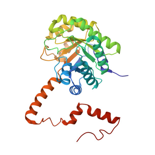
3FA3

Crystal structure of 2,3-dimethylmalate lyase, a PEP mutase/isocitrate lyase superfamily member, trigonal crystal form


Experimental Data Snapshot

  • Method: X-RAY DIFFRACTION
  • Resolution: 2.60 Å
  • R-Value Free: 0.255 
  • R-Value Work: 0.190 
  • R-Value Observed: 0.193 

wwPDB Validation   3D Report Full Report


Ligand Structure Quality Assessment 


This is version 1.2 of the entry. See complete history


Literature

Structure and function of 2,3-dimethylmalate lyase, a PEP mutase/isocitrate lyase superfamily member.

Narayanan, B.Niu, W.Joosten, H.J.Li, Z.Kuipers, R.K.Schaap, P.J.Dunaway-Mariano, D.Herzberg, O.

(2009) J Mol Biol 386: 486-503

  • DOI: https://doi.org/10.1016/j.jmb.2008.12.037
  • Primary Citation of Related Structures:  
    3FA3, 3FA4

  • PubMed Abstract: 

    The Aspergillus niger genome contains four genes that encode proteins exhibiting greater than 30% amino acid sequence identity to the confirmed oxaloacetate acetyl hydrolase (OAH), an enzyme that belongs to the phosphoenolpyruvate mutase/isocitrate lyase superfamily. Previous studies have shown that a mutant A. niger strain lacking the OAH gene does not produce oxalate. To identify the function of the protein sharing the highest amino acid sequence identity with the OAH (An07g08390, Swiss-Prot entry Q2L887, 57% identity), we produced the protein in Escherichia coli and purified it for structural and functional studies. A focused substrate screen was used to determine the catalytic function of An07g08390 as (2R,3S)-dimethylmalate lyase (DMML): k(cat)=19.2 s(-1) and K(m)=220 microM. DMML also possesses significant OAH activity (k(cat)=0.5 s(-1) and K(m) =220 microM). DNA array analysis showed that unlike the A. niger oah gene, the DMML encoding gene is subject to catabolite repression. DMML is a key enzyme in bacterial nicotinate catabolism, catalyzing the last of nine enzymatic steps. This pathway does not have a known fungal counterpart. BLAST analysis of the A. niger genome for the presence of a similar pathway revealed the presence of homologs to only some of the pathway enzymes. This and the finding that A. niger does not thrive on nicotinamide as a sole carbon source suggest that the fungal DMML functions in a presently unknown metabolic pathway. The crystal structure of A. niger DMML (in complex with Mg(2+) and in complex with Mg(2+) and a substrate analog: the gem-diol of 3,3-difluoro-oxaloacetate) was determined for the purpose of identifying structural determinants of substrate recognition and catalysis. Structure-guided site-directed mutants were prepared and evaluated to test the contributions made by key active-site residues. In this article, we report the results in the broader context of the lyase branch of the phosphoenolpyruvate mutase/isocitrate lyase superfamily to provide insight into the evolution of functional diversity.


  • Organizational Affiliation

    Center for Advanced Research in Biotechnology, University of Maryland Biotechnology Institute, Rockville, 20850, USA.


Macromolecules
Find similar proteins by:  (by identity cutoff)  |  3D Structure
Entity ID: 1
MoleculeChains Sequence LengthOrganismDetailsImage
2,3-dimethylmalate lyase
A, B, C, D, E
A, B, C, D, E, F, G, H, I, J, K, L, M, N, O, P
302Aspergillus nigerMutation(s): 0 
Gene Names: An07g08390
EC: 4.1.3.32
UniProt
Find proteins for Q2L887 (Aspergillus niger)
Explore Q2L887 
Go to UniProtKB:  Q2L887
Entity Groups  
Sequence Clusters30% Identity50% Identity70% Identity90% Identity95% Identity100% Identity
UniProt GroupQ2L887
Sequence Annotations
Expand
  • Reference Sequence
Small Molecules
Ligands 3 Unique
IDChains Name / Formula / InChI Key2D Diagram3D Interactions
OAF
Query on OAF

Download Ideal Coordinates CCD File 
BA [auth E]
EA [auth F]
GA [auth G]
JA [auth H]
LA [auth I]
BA [auth E],
EA [auth F],
GA [auth G],
JA [auth H],
LA [auth I],
OA [auth K],
QA [auth L],
R [auth A],
SA [auth M],
V [auth B],
WA [auth O],
X [auth C],
YA [auth P],
Z [auth D]
2,2-difluoro-3,3-dihydroxybutanedioic acid
C4 H4 F2 O6
PNUBNHHFDUCRFW-UHFFFAOYSA-N
GOL
Query on GOL

Download Ideal Coordinates CCD File 
CA [auth E],
HA [auth G],
S [auth A],
T [auth A],
TA [auth M]
GLYCEROL
C3 H8 O3
PEDCQBHIVMGVHV-UHFFFAOYSA-N
MN
Query on MN

Download Ideal Coordinates CCD File 
AA [auth E]
DA [auth F]
FA [auth G]
IA [auth H]
KA [auth I]
AA [auth E],
DA [auth F],
FA [auth G],
IA [auth H],
KA [auth I],
MA [auth J],
NA [auth K],
PA [auth L],
Q [auth A],
RA [auth M],
U [auth B],
UA [auth N],
VA [auth O],
W [auth C],
XA [auth P],
Y [auth D]
MANGANESE (II) ION
Mn
WAEMQWOKJMHJLA-UHFFFAOYSA-N
Binding Affinity Annotations 
IDSourceBinding Affinity
OAF PDBBind:  3FA3 Ki: 2500 (nM) from 1 assay(s)
Experimental Data & Validation

Experimental Data

  • Method: X-RAY DIFFRACTION
  • Resolution: 2.60 Å
  • R-Value Free: 0.255 
  • R-Value Work: 0.190 
  • R-Value Observed: 0.193 
  • Space Group: P 32
Unit Cell:
Length ( Å )Angle ( ˚ )
a = 160.57α = 90
b = 160.57β = 90
c = 161.41γ = 120
Software Package:
Software NamePurpose
CrystalCleardata collection
PHASERphasing
REFMACrefinement
CrystalCleardata reduction
CrystalCleardata scaling

Structure Validation

View Full Validation Report


Ligand Structure Quality Assessment 


Entry History 

Deposition Data

Revision History  (Full details and data files)

  • Version 1.0: 2009-01-27
    Type: Initial release
  • Version 1.1: 2011-07-13
    Changes: Advisory, Refinement description, Version format compliance
  • Version 1.2: 2023-12-27
    Changes: Data collection, Database references, Derived calculations