3F0Z

Crystal structure of Clostridium acetobutylicum 8-oxoguanine glycosylase/lyase in its apo-form


Experimental Data Snapshot

  • Method: X-RAY DIFFRACTION
  • Resolution: 2.20 Å
  • R-Value Free: 0.217 
  • R-Value Work: 0.174 
  • R-Value Observed: 0.174 

wwPDB Validation   3D Report Full Report


This is version 2.0 of the entry. See complete history


Literature

Structural characterization of Clostridium acetobutylicum 8-oxoguanine DNA glycosylase in its apo form and in complex with 8-oxodeoxyguanosine.

Faucher, F.Robey-Bond, S.M.Wallace, S.S.Doublie, S.

(2009) J Mol Biol 387: 669-679

  • DOI: https://doi.org/10.1016/j.jmb.2009.01.067
  • Primary Citation of Related Structures:  
    3F0Z, 3F10

  • PubMed Abstract: 

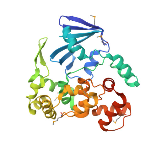
    DNA is subject to a multitude of oxidative damages generated by oxidizing agents from metabolism and exogenous sources and by ionizing radiation. Guanine is particularly vulnerable to oxidation, and the most common oxidative product 8-oxoguanine (8-oxoG) is the most prevalent lesion observed in DNA molecules. 8-OxoG can form a normal Watson-Crick pair with cytosine (8-oxoG:C), but it can also form a stable Hoogsteen pair with adenine (8-oxoG:A), leading to a G:C-->T:A transversion after replication. Fortunately, 8-oxoG is recognized and excised by either of two DNA glycosylases of the base excision repair pathway: formamidopyrimidine-DNA glycosylase and 8-oxoguanine DNA glycosylase (Ogg). While Clostridium acetobutylicum Ogg (CacOgg) DNA glycosylase can specifically recognize and remove 8-oxoG, it displays little preference for the base opposite the lesion, which is unusual for a member of the Ogg1 family. This work describes the crystal structures of CacOgg in its apo form and in complex with 8-oxo-2'-deoxyguanosine. A structural comparison between the apo form and the liganded form of the enzyme reveals a structural reorganization of the C-terminal domain upon binding of 8-oxoG, similar to that reported for human OGG1. A structural comparison of CacOgg with human OGG1, in complex with 8-oxoG containing DNA, provides a structural rationale for the lack of opposite base specificity displayed by CacOgg.


  • Organizational Affiliation

    Department of Microbiology and Molecular Genetics, The Markey Center for Molecular Genetics, University of Vermont, Stafford Hall, 95 Carrigan Drive, Burlington, VT 05405-0068, USA.


Macromolecules
Find similar proteins by:  (by identity cutoff)  |  3D Structure
Entity ID: 1
MoleculeChains Sequence LengthOrganismDetailsImage
8-oxoguanine-DNA-glycosylase292Clostridium acetobutylicumMutation(s): 0 
Gene Names: CAC2707CA_C2707
EC: 3.2.2 (PDB Primary Data), 4.2.99.18 (PDB Primary Data)
UniProt
Find proteins for Q97FM4 (Clostridium acetobutylicum (strain ATCC 824 / DSM 792 / JCM 1419 / LMG 5710 / VKM B-1787))
Explore Q97FM4 
Go to UniProtKB:  Q97FM4
Entity Groups  
Sequence Clusters30% Identity50% Identity70% Identity90% Identity95% Identity100% Identity
UniProt GroupQ97FM4
Sequence Annotations
Expand
  • Reference Sequence
Small Molecules
Ligands 1 Unique
IDChains Name / Formula / InChI Key2D Diagram3D Interactions
GOL
Query on GOL

Download Ideal Coordinates CCD File 
B [auth A]GLYCEROL
C3 H8 O3
PEDCQBHIVMGVHV-UHFFFAOYSA-N
Modified Residues  1 Unique
IDChains TypeFormula2D DiagramParent
MSE
Query on MSE
A
L-PEPTIDE LINKINGC5 H11 N O2 SeMET
Experimental Data & Validation

Experimental Data

  • Method: X-RAY DIFFRACTION
  • Resolution: 2.20 Å
  • R-Value Free: 0.217 
  • R-Value Work: 0.174 
  • R-Value Observed: 0.174 
  • Space Group: P 41 21 2
Unit Cell:
Length ( Å )Angle ( ˚ )
a = 61.35α = 90
b = 61.35β = 90
c = 158.9γ = 90
Software Package:
Software NamePurpose
SCALAdata scaling
SHARPphasing
CNSrefinement
PDB_EXTRACTdata extraction
IDBdata collection
XSCALEdata scaling

Structure Validation

View Full Validation Report


Entry History 

Deposition Data

Revision History  (Full details and data files)

  • Version 1.0: 2009-04-07
    Type: Initial release
  • Version 1.1: 2011-07-13
    Changes: Non-polymer description, Version format compliance
  • Version 1.2: 2012-03-21
    Changes: Database references
  • Version 2.0: 2023-12-27
    Changes: Atomic model, Data collection, Database references, Derived calculations