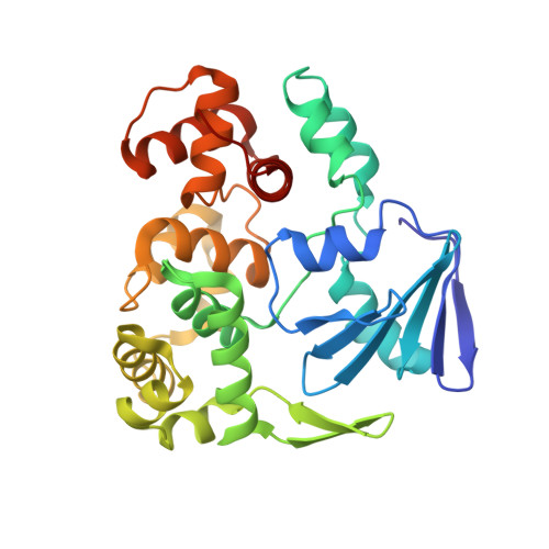
3F10

Crystal structure of Clostridium Acetobutylicum 8-oxoguanine DNA glycosylase in complex with 8-oxoguanosine


Experimental Data Snapshot

  • Method: X-RAY DIFFRACTION
  • Resolution: 2.30 Å
  • R-Value Free: 0.254 
  • R-Value Work: 0.215 
  • R-Value Observed: 0.215 

wwPDB Validation   3D Report Full Report


Ligand Structure Quality Assessment 


This is version 1.3 of the entry. See complete history


Literature

Structural characterization of Clostridium acetobutylicum 8-oxoguanine DNA glycosylase in its apo form and in complex with 8-oxodeoxyguanosine.

Faucher, F.Robey-Bond, S.M.Wallace, S.S.Doublie, S.

(2009) J Mol Biol 387: 669-679

  • DOI: 10.1016/j.jmb.2009.01.067
  • Primary Citation of Related Structures:  
    3F0Z, 3F10

  • PubMed Abstract: 
  • DNA is subject to a multitude of oxidative damages generated by oxidizing agents from metabolism and exogenous sources and by ionizing radiation. Guanine is particularly vulnerable to oxidation, and the most common oxidative product 8-oxoguanine (8-oxoG) is the most prevalent lesion observed in DNA molecules ...

    DNA is subject to a multitude of oxidative damages generated by oxidizing agents from metabolism and exogenous sources and by ionizing radiation. Guanine is particularly vulnerable to oxidation, and the most common oxidative product 8-oxoguanine (8-oxoG) is the most prevalent lesion observed in DNA molecules. 8-OxoG can form a normal Watson-Crick pair with cytosine (8-oxoG:C), but it can also form a stable Hoogsteen pair with adenine (8-oxoG:A), leading to a G:C-->T:A transversion after replication. Fortunately, 8-oxoG is recognized and excised by either of two DNA glycosylases of the base excision repair pathway: formamidopyrimidine-DNA glycosylase and 8-oxoguanine DNA glycosylase (Ogg). While Clostridium acetobutylicum Ogg (CacOgg) DNA glycosylase can specifically recognize and remove 8-oxoG, it displays little preference for the base opposite the lesion, which is unusual for a member of the Ogg1 family. This work describes the crystal structures of CacOgg in its apo form and in complex with 8-oxo-2'-deoxyguanosine. A structural comparison between the apo form and the liganded form of the enzyme reveals a structural reorganization of the C-terminal domain upon binding of 8-oxoG, similar to that reported for human OGG1. A structural comparison of CacOgg with human OGG1, in complex with 8-oxoG containing DNA, provides a structural rationale for the lack of opposite base specificity displayed by CacOgg.


    Organizational Affiliation

    Department of Microbiology and Molecular Genetics, The Markey Center for Molecular Genetics, University of Vermont, Stafford Hall, 95 Carrigan Drive, Burlington, VT 05405-0068, USA.


Macromolecules
Find similar proteins by:  (by identity cutoff)  |  3D Structure
Entity ID: 1
MoleculeChainsSequence LengthOrganismDetailsImage
8-oxoguanine-DNA-glycosylaseA292Clostridium acetobutylicumMutation(s): 1 
Gene Names: CAC2707CA_C2707
EC: 3.2.2 (PDB Primary Data), 4.2.99.18 (PDB Primary Data)
UniProt
Find proteins for Q97FM4 (Clostridium acetobutylicum (strain ATCC 824 / DSM 792 / JCM 1419 / LMG 5710 / VKM B-1787))
Explore Q97FM4 
Go to UniProtKB:  Q97FM4
Entity Groups  
Sequence Clusters30% Identity50% Identity70% Identity90% Identity95% Identity100% Identity
UniProt GroupQ97FM4
Protein Feature View
Expand
  • Reference Sequence
Small Molecules
Ligands 2 Unique
IDChainsName / Formula / InChI Key2D Diagram3D Interactions
8HG
Query on 8HG

Download Ideal Coordinates CCD File 
B [auth A]2'-DEOXY-8-OXOGUANOSINE
C10 H13 N5 O5
HCAJQHYUCKICQH-VPENINKCSA-N
 Ligand Interaction
EDO
Query on EDO

Download Ideal Coordinates CCD File 
C [auth A],
D [auth A]
1,2-ETHANEDIOL
C2 H6 O2
LYCAIKOWRPUZTN-UHFFFAOYSA-N
 Ligand Interaction
Experimental Data & Validation

Experimental Data

  • Method: X-RAY DIFFRACTION
  • Resolution: 2.30 Å
  • R-Value Free: 0.254 
  • R-Value Work: 0.215 
  • R-Value Observed: 0.215 
  • Space Group: P 41 3 2
Unit Cell:
Length ( Å )Angle ( ˚ )
a = 138.5α = 90
b = 138.5β = 90
c = 138.5γ = 90
Software Package:
Software NamePurpose
XSCALEdata scaling
MOLREPphasing
CNSrefinement
PDB_EXTRACTdata extraction
MAR345dtbdata collection
XDSdata reduction

Structure Validation

View Full Validation Report


Ligand Structure Quality Assessment 


Entry History 

Deposition Data

Revision History  (Full details and data files)

  • Version 1.0: 2009-04-07
    Type: Initial release
  • Version 1.1: 2011-07-13
    Changes: Version format compliance
  • Version 1.2: 2012-03-21
    Changes: Database references
  • Version 1.3: 2021-10-20
    Changes: Database references, Derived calculations