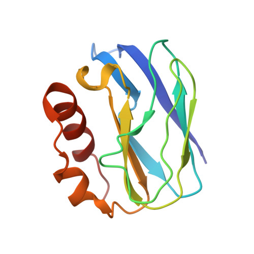
3EF4

Crystal structure of native pseudoazurin from Hyphomicrobium denitrificans


Experimental Data Snapshot

  • Method: X-RAY DIFFRACTION
  • Resolution: 1.18 Å
  • R-Value Free: 0.169 
  • R-Value Work: 0.135 
  • R-Value Observed: 0.135 

Starting Model: experimental
View more details

wwPDB Validation   3D Report Full Report


This is version 1.2 of the entry. See complete history


Literature

Atomic resolution structure of pseudoazurin from the methylotrophic denitrifying bacterium Hyphomicrobium denitrificans: structural insights into its spectroscopic properties

Hira, D.Nojiri, M.Suzuki, S.

(2009) Acta Crystallogr D Biol Crystallogr 65: 85-92

  • DOI: https://doi.org/10.1107/S0907444908040195
  • Primary Citation of Related Structures:  
    3EF4

  • PubMed Abstract: 

    The crystal structure of native pseudoazurin (HdPAz) from the methylotrophic denitrifying bacterium Hyphomicrobium denitrificans has been determined at a resolution of 1.18 A. After refinement with SHELX employing anisotropic displacement parameters and riding H atoms, R(work) and R(free) were 0.135 and 0.169, respectively. Visualization of the anisotropic displacement parameters as thermal ellipsoids provided insight into the atomic motion within the perturbed type 1 Cu site. The asymmetric unit includes three HdPAz molecules which are tightly packed by head-to-head cupredoxin dimer formation. The shape of the Cu-atom ellipsoid implies significant vibrational motion diagonal to the equatorial xy plane defined by the three ligands (two His and one Cys). The geometric parameters of the type 1 Cu site in the HdPAz structure differ unambiguously from those of other pseudoazurins. It is demonstrated that their structural aspects are consistent with the unique visible absorption spectrum.


  • Organizational Affiliation

    Bioinorganic Chemistry Laboratory, Department of Chemistry, Graduate School of Science, Osaka University, 1-1 Machikaneyama, Toyonaka, Osaka 560-0043, Japan.


Macromolecules
Find similar proteins by:  (by identity cutoff)  |  3D Structure
Entity ID: 1
MoleculeChains Sequence LengthOrganismDetailsImage
Blue copper protein
A, B, C
124Hyphomicrobium denitrificansMutation(s): 0 
UniProt
Find proteins for A7VL37 (Hyphomicrobium denitrificans)
Explore A7VL37 
Go to UniProtKB:  A7VL37
Entity Groups  
Sequence Clusters30% Identity50% Identity70% Identity90% Identity95% Identity100% Identity
UniProt GroupA7VL37
Sequence Annotations
Expand
  • Reference Sequence
Small Molecules
Ligands 2 Unique
IDChains Name / Formula / InChI Key2D Diagram3D Interactions
PO4
Query on PO4

Download Ideal Coordinates CCD File 
E [auth A]
F [auth A]
G [auth A]
H [auth A]
J [auth B]
E [auth A],
F [auth A],
G [auth A],
H [auth A],
J [auth B],
K [auth B],
L [auth B],
M [auth B],
N [auth B],
O [auth B],
Q [auth C],
R [auth C],
S [auth C],
T [auth C],
U [auth C]
PHOSPHATE ION
O4 P
NBIIXXVUZAFLBC-UHFFFAOYSA-K
CU
Query on CU

Download Ideal Coordinates CCD File 
D [auth A],
I [auth B],
P [auth C]
COPPER (II) ION
Cu
JPVYNHNXODAKFH-UHFFFAOYSA-N
Experimental Data & Validation

Experimental Data

  • Method: X-RAY DIFFRACTION
  • Resolution: 1.18 Å
  • R-Value Free: 0.169 
  • R-Value Work: 0.135 
  • R-Value Observed: 0.135 
  • Space Group: C 1 2 1
Unit Cell:
Length ( Å )Angle ( ˚ )
a = 97.69α = 90
b = 50.623β = 90.98
c = 82.378γ = 90
Software Package:
Software NamePurpose
HKL-2000data collection
MOLREPphasing
SHELXL-97refinement
HKL-2000data reduction
SCALEPACKdata scaling

Structure Validation

View Full Validation Report


Entry History 

Deposition Data

Revision History  (Full details and data files)

  • Version 1.0: 2008-12-30
    Type: Initial release
  • Version 1.1: 2011-07-13
    Changes: Version format compliance
  • Version 1.2: 2023-11-01
    Changes: Data collection, Database references, Derived calculations, Refinement description