3EAB

Crystal structure of Spastin MIT in complex with ESCRT III


Experimental Data Snapshot

  • Method: X-RAY DIFFRACTION
  • Resolution: 2.50 Å
  • R-Value Free: 0.268 
  • R-Value Work: 0.232 
  • R-Value Observed: 0.232 

wwPDB Validation   3D Report Full Report


This is version 1.2 of the entry. See complete history


Literature

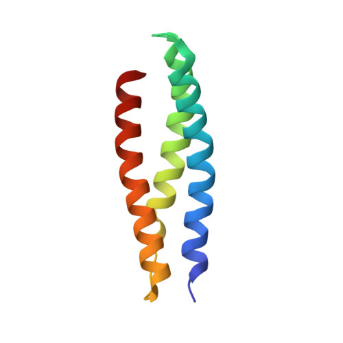
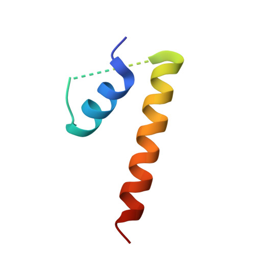
Structural basis for midbody targeting of spastin by the ESCRT-III protein CHMP1B.

Yang, D.Rismanchi, N.Renvoise, B.Lippincott-Schwartz, J.Blackstone, C.Hurley, J.H.

(2008) Nat Struct Mol Biol 15: 1278-1286

  • DOI: https://doi.org/10.1038/nsmb.1512
  • Primary Citation of Related Structures:  
    3EAB

  • PubMed Abstract: 

    The endosomal sorting complex required for transport (ESCRT) machinery, including ESCRT-III, localizes to the midbody and participates in the membrane-abscission step of cytokinesis. The ESCRT-III protein charged multivesicular body protein 1B (CHMP1B) is required for recruitment of the MIT domain-containing protein spastin, a microtubule-severing enzyme, to the midbody. The 2.5-A structure of the C-terminal tail of CHMP1B with the MIT domain of spastin reveals a specific, high-affinity complex involving a noncanonical binding site between the first and third helices of the MIT domain. The structural interface is twice as large as that of the MIT domain of the VPS4-CHMP complex, consistent with the high affinity of the interaction. A series of unique hydrogen-bonding interactions and close packing of small side chains discriminate against the other ten human ESCRT-III subunits. Point mutants in the CHMP1B binding site of spastin block recruitment of spastin to the midbody and impair cytokinesis.


  • Organizational Affiliation

    Laboratory of Molecular Biology, National Institute of Diabetes and Digestive and Kidney Diseases, National Institutes of Health, US Department of Health and Human Services, Bethesda, Maryland 20892, USA.


Macromolecules
Find similar proteins by:  (by identity cutoff)  |  3D Structure
Entity ID: 1
MoleculeChains Sequence LengthOrganismDetailsImage
Spastin
A, B, C, D, E
A, B, C, D, E, F
89Homo sapiensMutation(s): 0 
Gene Names: SPASTKIAA1083SPG4
UniProt & NIH Common Fund Data Resources
Find proteins for Q9UBP0 (Homo sapiens)
Explore Q9UBP0 
Go to UniProtKB:  Q9UBP0
PHAROS:  Q9UBP0
GTEx:  ENSG00000021574 
Entity Groups  
Sequence Clusters30% Identity50% Identity70% Identity90% Identity95% Identity100% Identity
UniProt GroupQ9UBP0
Sequence Annotations
Expand
  • Reference Sequence
Find similar proteins by:  (by identity cutoff)  |  3D Structure
Entity ID: 2
MoleculeChains Sequence LengthOrganismDetailsImage
CHMP1b
G, H, I, J, K
G, H, I, J, K, L
50Homo sapiensMutation(s): 0 
UniProt & NIH Common Fund Data Resources
Find proteins for Q7LBR1 (Homo sapiens)
Explore Q7LBR1 
Go to UniProtKB:  Q7LBR1
PHAROS:  Q7LBR1
GTEx:  ENSG00000255112 
Entity Groups  
Sequence Clusters30% Identity50% Identity70% Identity90% Identity95% Identity100% Identity
UniProt GroupQ7LBR1
Sequence Annotations
Expand
  • Reference Sequence
Experimental Data & Validation

Experimental Data

  • Method: X-RAY DIFFRACTION
  • Resolution: 2.50 Å
  • R-Value Free: 0.268 
  • R-Value Work: 0.232 
  • R-Value Observed: 0.232 
  • Space Group: P 21 21 2
Unit Cell:
Length ( Å )Angle ( ˚ )
a = 151.967α = 90
b = 95.493β = 90
c = 100.36γ = 90
Software Package:
Software NamePurpose
SOLVEphasing
CNSrefinement
REFMACrefinement
HKL-2000data reduction
HKL-2000data scaling

Structure Validation

View Full Validation Report


Entry History 

Deposition Data

Revision History  (Full details and data files)

  • Version 1.0: 2008-11-11
    Type: Initial release
  • Version 1.1: 2011-07-13
    Changes: Version format compliance
  • Version 1.2: 2024-02-21
    Changes: Data collection, Database references