3E6O | pdb_00003e6o

Structure of murine INOS oxygenase domain with inhibitor AR-C124355


Experimental Data Snapshot

  • Method: X-RAY DIFFRACTION
  • Resolution: 2.60 Å
  • R-Value Free: 
    0.262 (Depositor), 0.436 (DCC) 
  • R-Value Work: 
    0.229 (Depositor), 0.416 (DCC) 

wwPDB Validation 3D Report Full Report

Validation slider image for 3E6O

Ligand Structure Quality Assessment 


This is version 1.3 of the entry. See complete history

Literature

Anchored plasticity opens doors for selective inhibitor design in nitric oxide synthase.

Garcin, E.D.Arvai, A.S.Rosenfeld, R.J.Kroeger, M.D.Crane, B.R.Andersson, G.Andrews, G.Hamley, P.J.Mallinder, P.R.Nicholls, D.J.St-Gallay, S.A.Tinker, A.C.Gensmantel, N.P.Mete, A.Cheshire, D.R.Connolly, S.Stuehr, D.J.Aberg, A.Wallace, A.V.Tainer, J.A.Getzoff, E.D.

(2008) Nat Chem Biol 4: 700-707

  • DOI: https://doi.org/10.1038/nchembio.115
  • Primary Citation Related Structures: 
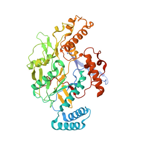
    3E65, 3E67, 3E68, 3E6L, 3E6N, 3E6O, 3E6T, 3E7G, 3E7I, 3E7M, 3E7S, 3E7T, 3EAH, 3EAI, 3EBD, 3EBF, 3EJ8

  • PubMed Abstract: 

    Nitric oxide synthase (NOS) enzymes synthesize nitric oxide, a signal for vasodilatation and neurotransmission at low concentrations and a defensive cytotoxin at higher concentrations. The high active site conservation among all three NOS isozymes hinders the design of selective NOS inhibitors to treat inflammation, arthritis, stroke, septic shock and cancer. Our crystal structures and mutagenesis results identified an isozyme-specific induced-fit binding mode linking a cascade of conformational changes to a new specificity pocket. Plasticity of an isozyme-specific triad of distant second- and third-shell residues modulates conformational changes of invariant first-shell residues to determine inhibitor selectivity. To design potent and selective NOS inhibitors, we developed the anchored plasticity approach: anchor an inhibitor core in a conserved binding pocket, then extend rigid bulky substituents toward remote specificity pockets, which become accessible upon conformational changes of flexible residues. This approach exemplifies general principles for the design of selective enzyme inhibitors that overcome strong active site conservation.


  • Organizational Affiliation
    • The Scripps Research Institute, Department of Molecular Biology and Skaggs Institute for Chemical Biology, 10550 North Torrey Pines Road, MB4, La Jolla, California 92037, USA. egarcin@umbc.edu

Macromolecule Content 

  • Total Structure Weight: 102.6 kDa 
  • Atom Count: 7,171 
  • Modeled Residue Count: 825 
  • Deposited Residue Count: 866 
  • Unique protein chains: 1

Macromolecules

Find similar proteins by:|  3D Structure
Entity ID: 1
MoleculeChains  Sequence LengthOrganismDetailsImage
Nitric oxide synthase, inducible
A, B
433Mus musculusMutation(s): 0 
EC: 1.14.13.39
UniProt & NIH Common Fund Data Resources
Find proteins for P29477 (Mus musculus)
Explore P29477 
Go to UniProtKB:  P29477
IMPC:  MGI:97361
Entity Groups
Sequence Clusters30% Identity50% Identity70% Identity90% Identity95% Identity100% Identity
UniProt GroupP29477
Sequence Annotations
Expand
Reference Sequence

Small Molecules

Ligands 3 Unique
IDChains Name / Formula / InChI Key2D Diagram3D Interactions
HEM

Query on HEM


Download:Ideal Coordinates CCD File
C [auth A],
F [auth B]
PROTOPORPHYRIN IX CONTAINING FE
C34 H32 Fe N4 O4
KABFMIBPWCXCRK-RGGAHWMASA-L
A55

Query on A55


Download:Ideal Coordinates CCD File
E [auth A],
H [auth B]
N-[2-(4-AMINO-5,8-DIFLUORO-1,2-DIHYDROQUINAZOLIN-2-YL)ETHYL]-3-FURAMIDE
C15 H14 F2 N4 O2
BOAUWUUBSXECNL-LLVKDONJSA-N
H4B

Query on H4B


Download:Ideal Coordinates CCD File
D [auth A],
G [auth B]
5,6,7,8-TETRAHYDROBIOPTERIN
C9 H15 N5 O3
FNKQXYHWGSIFBK-RPDRRWSUSA-N
Binding Affinity Annotations 
IDSourceBinding Affinity
A55 BindingDB:  3E6O IC50: 31 (nM) from 1 assay(s)

Experimental Data & Validation

Experimental Data

  • Method: X-RAY DIFFRACTION
  • Resolution: 2.60 Å
  • R-Value Free:  0.262 (Depositor), 0.436 (DCC) 
  • R-Value Work:  0.229 (Depositor), 0.416 (DCC) 
Space Group: P 61 2 2
Unit Cell:
Length ( Å )Angle ( ˚ )
a = 213.898α = 90
b = 213.898β = 90
c = 116.148γ = 120
Software Package:
Software NamePurpose
CNSrefinement
PDB_EXTRACTdata extraction
DENZOdata reduction
SCALEPACKdata scaling

Structure Validation

View Full Validation Report


Ligand Structure Quality Assessment 


Entry History 

Revision History  (Full details and data files)

  • Version 1.0: 2008-10-07
    Type: Initial release
  • Version 1.1: 2011-07-13
    Changes: Version format compliance
  • Version 1.2: 2017-10-25
    Changes: Refinement description
  • Version 1.3: 2024-02-21
    Changes: Data collection, Database references, Derived calculations