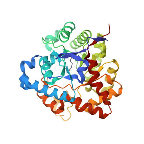
Balancing the stability and the catalytic specificities of OP hydrolases with enhanced V-agent activities.
Reeves, T.E., Wales, M.E., Grimsley, J.K., Li, P., Cerasoli, D.M., Wild, J.R.(2008) Protein Eng Des Sel 21: 405-412
- PubMed: 18434422
- DOI: https://doi.org/10.1093/protein/gzn019
- Primary Citation of Related Structures:
3E3H - PubMed Abstract:
Rational site-directed mutagenesis and biophysical analyses have been used to explore the thermodynamic stability and catalytic capabilities of organophosphorus hydrolase (OPH) and its genetically modified variants. There are clear trade-offs in the stability of modifications that enhance catalytic activities. For example, the H254R/H257L variant has higher turnover numbers for the chemical warfare agents VX (144 versus 14 s(-1) for the native enzyme (wild type) and VR (Russian VX, 465 versus 12 s(-1) for wild type). These increases are accompanied by a loss in stability in which the total Gibb's free energy for unfolding is 19.6 kcal/mol, which is 5.7 kcal/mol less than that of the wild-type enzyme. X-ray crystallographic studies support biophysical data that suggest amino acid residues near the active site contribute to the chemical and thermal stability through hydrophobic and cation-pi interactions. The cation-pi interactions appear to contribute an additional 7 kcal/mol to the overall global stability of the enzyme. Using rational design, it has been possible to make amino acid changes in this region that restored the stability, yet maintained effective V-agent activities, with turnover numbers of 68 and 36 s(-1) for VX and VR, respectively. This study describes the first rationally designed, stability/activity balance for an OPH enzyme with a legitimate V-agent activity, and its crystal structure.
- Department of Biochemistry and Biophysics, TAMU 2128, Texas A&M University, College Station, TX 77843-2128, USA.
Organizational Affiliation:


