3DVA

Snapshots of catalysis in the E1 subunit of the pyruvate dehydrogenase multi-enzyme complex


Experimental Data Snapshot

  • Method: X-RAY DIFFRACTION
  • Resolution: 2.35 Å
  • R-Value Free: 0.247 
  • R-Value Work: 0.190 
  • R-Value Observed: 0.192 

wwPDB Validation   3D Report Full Report


Ligand Structure Quality Assessment 


This is version 1.2 of the entry. See complete history


Literature

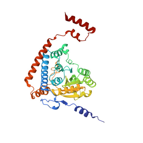
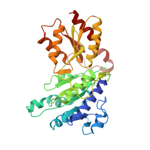
Snapshots of catalysis in the e1 subunit of the pyruvate dehydrogenase multienzyme complex

Pei, X.Y.Titman, C.M.Frank, R.A.Leeper, F.J.Luisi, B.F.

(2008) Structure 16: 1860-1872

  • DOI: https://doi.org/10.1016/j.str.2008.10.009
  • Primary Citation of Related Structures:  
    3DUF, 3DV0, 3DVA

  • PubMed Abstract: 
  • The pyruvate dehydrogenase multienzyme assembly (PDH) generates acetyl coenzyme A and reducing equivalents from pyruvate in a multiple-step process that is a nexus of central metabolism. We report crystal structures of the Geobacillus stearothermophilus PDH E1p subunit with ligands that mimic the prereaction complex and the postdecarboxylation product ...

    The pyruvate dehydrogenase multienzyme assembly (PDH) generates acetyl coenzyme A and reducing equivalents from pyruvate in a multiple-step process that is a nexus of central metabolism. We report crystal structures of the Geobacillus stearothermophilus PDH E1p subunit with ligands that mimic the prereaction complex and the postdecarboxylation product. The structures implicate residues that help to orient substrates, nurture intermediates, and organize surface loops so that they can engage a mobile lipoyl domain that receives the acetyl group and shuttles it to the next active site. The structural and enzymatic data suggest that H128beta performs a dual role: first, as electrostatic catalyst of the reaction of pyruvate with the thiamine cofactor; and second, as a proton donor in the second reaction of acetyl group with the lipoate. We also identify I206alpha as a key residue in mediating the conformation of active-site loops. We propose that a simple conformational flip of the H271alpha side chain assists transfer of the acetyl group from thiamine cofactor to lipoyl domain in synchrony with reduction of the dithiolane ring.


    Organizational Affiliation

    Department of Biochemistry, University of Cambridge, 80 Tennis Court Road, Cambridge CB2 1GA, UK.


Macromolecules
Find similar proteins by:  (by identity cutoff)  |  3D Structure
Entity ID: 1
MoleculeChainsSequence LengthOrganismDetailsImage
Pyruvate dehydrogenase E1 component subunit alphaA, C, E, G369Geobacillus stearothermophilusMutation(s): 1 
Gene Names: pdhA
EC: 1.2.4.1
UniProt
Find proteins for P21873 (Geobacillus stearothermophilus)
Explore P21873 
Go to UniProtKB:  P21873
Entity Groups  
Sequence Clusters30% Identity50% Identity70% Identity90% Identity95% Identity100% Identity
UniProt GroupP21873
Protein Feature View
Expand
  • Reference Sequence
Find similar proteins by:  (by identity cutoff)  |  3D Structure
Entity ID: 2
MoleculeChainsSequence LengthOrganismDetailsImage
Pyruvate dehydrogenase E1 component subunit betaB, D, F, H325Geobacillus stearothermophilusMutation(s): 0 
Gene Names: pdhB
EC: 1.2.4.1
UniProt
Find proteins for P21874 (Geobacillus stearothermophilus)
Explore P21874 
Go to UniProtKB:  P21874
Entity Groups  
Sequence Clusters30% Identity50% Identity70% Identity90% Identity95% Identity100% Identity
UniProt GroupP21874
Protein Feature View
Expand
  • Reference Sequence
Find similar proteins by:  (by identity cutoff)  |  3D Structure
Entity ID: 3
MoleculeChainsSequence LengthOrganismDetailsImage
Dihydrolipoyllysine-residue acetyltransferase component of pyruvate dehydrogenase complexI, J428Geobacillus stearothermophilusMutation(s): 0 
Gene Names: pdhC
EC: 2.3.1.12
UniProt
Find proteins for P11961 (Geobacillus stearothermophilus)
Explore P11961 
Go to UniProtKB:  P11961
Entity Groups  
Sequence Clusters30% Identity50% Identity70% Identity90% Identity95% Identity100% Identity
UniProt GroupP11961
Protein Feature View
Expand
  • Reference Sequence
Small Molecules
Experimental Data & Validation

Experimental Data

  • Method: X-RAY DIFFRACTION
  • Resolution: 2.35 Å
  • R-Value Free: 0.247 
  • R-Value Work: 0.190 
  • R-Value Observed: 0.192 
  • Space Group: P 1 21 1
Unit Cell:
Length ( Å )Angle ( ˚ )
a = 68.673α = 90
b = 232.294β = 91.2
c = 91.936γ = 90
Software Package:
Software NamePurpose
REFMACrefinement
ADSCdata collection
MOSFLMdata reduction
SCALAdata scaling
AMoREphasing

Structure Validation

View Full Validation Report


Ligand Structure Quality Assessment 


Entry History 

Deposition Data

Revision History  (Full details and data files)

  • Version 1.0: 2009-01-13
    Type: Initial release
  • Version 1.1: 2011-07-13
    Changes: Version format compliance
  • Version 1.2: 2021-10-20
    Changes: Database references, Derived calculations, Source and taxonomy