3DSX

Crystal structure of RabGGTase(DELTA LRR; DELTA IG)in complex with di-prenylated peptide Ser-Cys(GG)-Ser-Cys(GG) derivated from Rab7


Experimental Data Snapshot

  • Method: X-RAY DIFFRACTION
  • Resolution: 2.10 Å
  • R-Value Free: 0.229 
  • R-Value Work: 0.168 
  • R-Value Observed: 0.171 

wwPDB Validation   3D Report Full Report


Ligand Structure Quality Assessment 


This is version 1.2 of the entry. See complete history


Literature

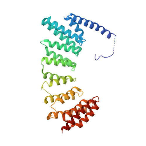
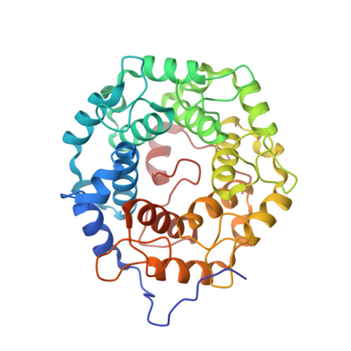
Structures of RabGGTase-substrate/product complexes provide insights into the evolution of protein prenylation

Guo, Z.Wu, Y.-W.Das, D.Delon, C.Cramer, J.Yu, S.Thuns, S.Lupilova, N.Waldmann, H.Brunsveld, L.Goody, R.S.Alexandrov, K.Blankenfeldt, W.

(2008) EMBO J 27: 2444-2456

  • DOI: https://doi.org/10.1038/emboj.2008.164
  • Primary Citation of Related Structures:  
    3DSS, 3DST, 3DSU, 3DSV, 3DSW, 3DSX

  • PubMed Abstract: 
  • Post-translational isoprenylation of proteins is carried out by three related enzymes: farnesyltransferase, geranylgeranyl transferase-I, and Rab geranylgeranyl transferase (RabGGTase). Despite the fact that the last one is responsible for the largest number of individual protein prenylation events in the cell, no structural information is available on its interaction with substrates and products ...

    Post-translational isoprenylation of proteins is carried out by three related enzymes: farnesyltransferase, geranylgeranyl transferase-I, and Rab geranylgeranyl transferase (RabGGTase). Despite the fact that the last one is responsible for the largest number of individual protein prenylation events in the cell, no structural information is available on its interaction with substrates and products. Here, we present structural and biophysical analyses of RabGGTase in complex with phosphoisoprenoids as well as with the prenylated peptides that mimic the C terminus of Rab7 GTPase. The data demonstrate that, unlike other protein prenyl transferases, both RabGGTase and its substrate RabGTPases completely 'outsource' their specificity for each other to an accessory subunit, the Rab escort protein (REP). REP mediates the placement of the C terminus of RabGTPase into the active site of RabGGTase through a series protein-protein interactions of decreasing strength and selectivity. This arrangement enables RabGGTase to prenylate any cysteine-containing sequence. On the basis of our structural and thermodynamic data, we propose that RabGGTase has evolved from a GGTase-I-like molecule that 'learned' to interact with a recycling factor (GDI) that, in turn, eventually gave rise to REP.


    Organizational Affiliation

    Department of Physical Biochemistry, Max-Planck-Institute for Molecular Physiology, Dortmund, Germany.


Macromolecules
Find similar proteins by:  (by identity cutoff)  |  3D Structure
Entity ID: 1
MoleculeChainsSequence LengthOrganismDetailsImage
Geranylgeranyl transferase type-2 subunit alphaA331Rattus norvegicusMutation(s): 0 
Gene Names: RabggtaGgta
EC: 2.5.1.60
UniProt
Find proteins for Q08602 (Rattus norvegicus)
Explore Q08602 
Go to UniProtKB:  Q08602
Entity Groups  
Sequence Clusters30% Identity50% Identity70% Identity90% Identity95% Identity100% Identity
UniProt GroupQ08602
Protein Feature View
Expand
  • Reference Sequence
Find similar proteins by:  (by identity cutoff)  |  3D Structure
Entity ID: 2
MoleculeChainsSequence LengthOrganismDetailsImage
Geranylgeranyl transferase type-2 subunit betaB331Rattus norvegicusMutation(s): 0 
Gene Names: RabggtbGgtb
EC: 2.5.1.60
UniProt
Find proteins for Q08603 (Rattus norvegicus)
Explore Q08603 
Go to UniProtKB:  Q08603
Entity Groups  
Sequence Clusters30% Identity50% Identity70% Identity90% Identity95% Identity100% Identity
UniProt GroupQ08603
Protein Feature View
Expand
  • Reference Sequence
Small Molecules
Ligands 3 Unique
IDChainsName / Formula / InChI Key2D Diagram3D Interactions
GER
Query on GER

Download Ideal Coordinates CCD File 
E [auth B]GERAN-8-YL GERAN
C20 H34
HSOYJGBJQAKCNA-CAIKYXSQSA-N
 Ligand Interaction
ZN
Query on ZN

Download Ideal Coordinates CCD File 
C [auth B]ZINC ION
Zn
PTFCDOFLOPIGGS-UHFFFAOYSA-N
 Ligand Interaction
CA
Query on CA

Download Ideal Coordinates CCD File 
D [auth B]CALCIUM ION
Ca
BHPQYMZQTOCNFJ-UHFFFAOYSA-N
 Ligand Interaction
Experimental Data & Validation

Experimental Data

  • Method: X-RAY DIFFRACTION
  • Resolution: 2.10 Å
  • R-Value Free: 0.229 
  • R-Value Work: 0.168 
  • R-Value Observed: 0.171 
  • Space Group: P 21 21 21
Unit Cell:
Length ( Å )Angle ( ˚ )
a = 67.354α = 90
b = 91.153β = 90
c = 114.591γ = 90
Software Package:
Software NamePurpose
REFMACrefinement
XDSdata reduction
XSCALEdata scaling
PHASERphasing

Structure Validation

View Full Validation Report


Ligand Structure Quality Assessment 


Entry History 

Deposition Data

Revision History  (Full details and data files)

  • Version 1.0: 2008-09-09
    Type: Initial release
  • Version 1.1: 2011-07-13
    Changes: Version format compliance
  • Version 1.2: 2017-08-09
    Changes: Source and taxonomy