


Crystal structure of CYP2R1 in complex with 1-alpha-hydroxy-vitamin D2.
Strushkevich, N.V., Tempel, W., Gilep, A.A., Loppnau, P., Arrowsmith, C.H., Edwards, A.M., Bountra, C., Wilkstrom, M., Bochkarev, A., Park, H.To be published.
Experimental Data Snapshot
Starting Model: experimental
View more details
Entity ID: 1 | |||||
|---|---|---|---|---|---|
| Molecule | Chains | Sequence Length | Organism | Details | Image |
| Cytochrome P450 2R1 | 479 | Homo sapiens | Mutation(s): 0 Gene Names: CYP2R1 EC: 1.14.14 (PDB Primary Data), 1.14.14.24 (UniProt) | 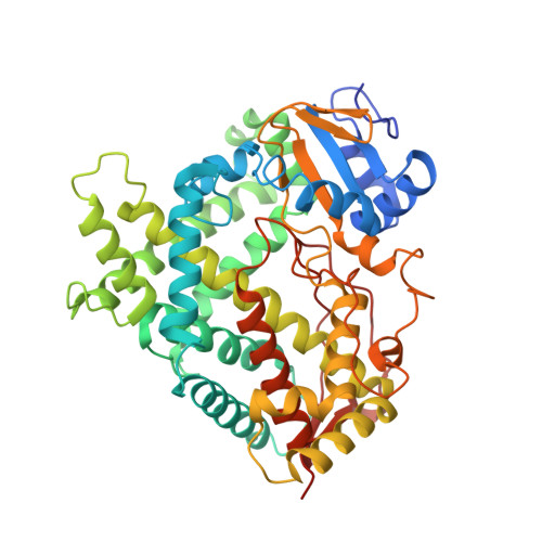 | |
UniProt & NIH Common Fund Data Resources | |||||
PHAROS: Q6VVX0 GTEx: ENSG00000186104 | |||||
Entity Groups | |||||
| Sequence Clusters | 30% Identity50% Identity70% Identity90% Identity95% Identity100% Identity | ||||
| UniProt Group | Q6VVX0 | ||||
Sequence AnnotationsExpand | |||||
Reference Sequence | |||||
| Ligands 2 Unique | |||||
|---|---|---|---|---|---|
| ID | Chains | Name / Formula / InChI Key | 2D Diagram | 3D Interactions | |
| HEM Download:Ideal Coordinates CCD File | E [auth A], G [auth B] | PROTOPORPHYRIN IX CONTAINING FE C34 H32 Fe N4 O4 KABFMIBPWCXCRK-RGGAHWMASA-L |  | ||
| V2H Download:Ideal Coordinates CCD File | F [auth A], H [auth B] | (1S,3R,5Z,7E,22E)-9,10-secoergosta-5,7,10,22-tetraene-1,3-diol C28 H44 O2 HKXBNHCUPKIYDM-CGMHZMFXSA-N |  | ||
Entity ID: 2 | |||||
|---|---|---|---|---|---|
| ID | Chains | Name | Type/Class | 2D Diagram | 3D Interactions |
| PRD_900012 Query on PRD_900012 | C, D | beta-cyclodextrin | Oligosaccharide / Drug delivery |  |
| Length ( Å ) | Angle ( ˚ ) |
|---|---|
| a = 137.303 | α = 90 |
| b = 163.04 | β = 90 |
| c = 152.588 | γ = 90 |
| Software Name | Purpose |
|---|---|
| DENZO | data reduction |
| SCALEPACK | data scaling |
| REFMAC | refinement |
| PDB_EXTRACT | data extraction |
| ADSC | data collection |
| HKL-3000 | data reduction |
| REFMAC | phasing |


