3D7A

Crystal structure of DUF54 family protein PH1010 from hyperthermophilic archaea Pyrococcus horikoshii OT3


Experimental Data Snapshot

  • Method: X-RAY DIFFRACTION
  • Resolution: 1.90 Å
  • R-Value Free: 0.281 
  • R-Value Work: 0.238 
  • R-Value Observed: 0.240 

wwPDB Validation   3D Report Full Report


This is version 1.3 of the entry. See complete history


Literature

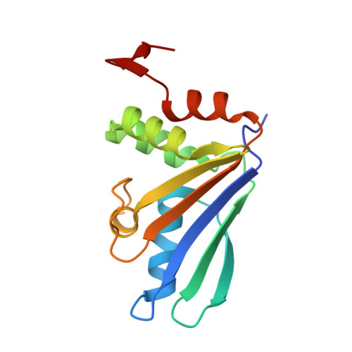
Crystal structure of the DUF54 family protein PH1010 from hyperthermophilic archaea Pyrococcus horikoshii OT3.

Miyazono, K.I.Shirokane, M.Sawano, Y.Tanokura, M.

(2008) Proteins 74: 256-260


Macromolecules
Find similar proteins by:  (by identity cutoff)  |  3D Structure
Entity ID: 1
MoleculeChains Sequence LengthOrganismDetailsImage
UPF0201 protein PH1010
A, B
138Pyrococcus horikoshiiMutation(s): 0 
Gene Names: PH1010
UniProt
Find proteins for O58738 (Pyrococcus horikoshii (strain ATCC 700860 / DSM 12428 / JCM 9974 / NBRC 100139 / OT-3))
Explore O58738 
Go to UniProtKB:  O58738
Entity Groups  
Sequence Clusters30% Identity50% Identity70% Identity90% Identity95% Identity100% Identity
UniProt GroupO58738
Sequence Annotations
Expand
  • Reference Sequence
Experimental Data & Validation

Experimental Data

  • Method: X-RAY DIFFRACTION
  • Resolution: 1.90 Å
  • R-Value Free: 0.281 
  • R-Value Work: 0.238 
  • R-Value Observed: 0.240 
  • Space Group: P 21 21 21
Unit Cell:
Length ( Å )Angle ( ˚ )
a = 46.934α = 90
b = 49.526β = 90
c = 132.695γ = 90
Software Package:
Software NamePurpose
DENZOdata reduction
SCALEPACKdata scaling
REFMACrefinement
PDB_EXTRACTdata extraction

Structure Validation

View Full Validation Report


Entry History 

Deposition Data

Revision History  (Full details and data files)

  • Version 1.0: 2008-12-09
    Type: Initial release
  • Version 1.1: 2011-07-13
    Changes: Version format compliance
  • Version 1.2: 2017-10-25
    Changes: Advisory, Refinement description
  • Version 1.3: 2024-03-20
    Changes: Advisory, Data collection, Database references