3D4Y | pdb_00003d4y

GOLGI MANNOSIDASE II complex with mannoimidazole


Experimental Data Snapshot

  • Method: X-RAY DIFFRACTION
  • Resolution: 1.52 Å
  • R-Value Free: 
    0.208 (Depositor), 0.188 (DCC) 
  • R-Value Work: 
    0.186 (Depositor), 0.184 (DCC) 
  • R-Value Observed: 
    0.186 (Depositor) 

wwPDB Validation 3D Report Full Report

Validation slider image for 3D4Y

Ligand Structure Quality Assessment 


This is version 1.5 of the entry. See complete history

Literature

Structural analysis of Golgi alpha-mannosidase II inhibitors identified from a focused glycosidase inhibitor screen.

Kuntz, D.A.Tarling, C.A.Withers, S.G.Rose, D.R.

(2008) Biochemistry 47: 10058-10068

  • DOI: https://doi.org/10.1021/bi8010785
  • Primary Citation Related Structures: 
    3D4Y, 3D4Z, 3D50, 3D51, 3D52

  • PubMed Abstract: 

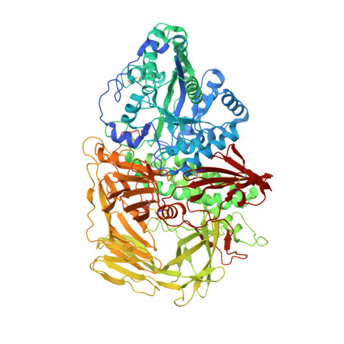
    The N-glycosylation pathway is a target for pharmaceutical intervention in a number of pathological conditions including cancer. Golgi alpha-mannosidase II (GMII) is the final glycoside hydrolase in the pathway and has been the target for a number of synthetic efforts aimed at providing more selective and effective inhibitors. Drosophila GMII (dGMII) has been extensively studied due to the ease of obtaining high resolution structural data, allowing the observation of substrate distortion upon binding and after formation of a trapped covalent reaction intermediate. However, attempts to find new inhibitor leads by high-throughput screening of large commercial libraries or through in silico docking were unsuccessful. In this paper we provide a kinetic and structural analysis of five inhibitors derived from a small glycosidase-focused library. Surprisingly, four of these were known inhibitors of beta-glucosidases. X-ray crystallographic analysis of the dGMII:inhibitor complexes highlights the ability of the zinc-containing GMII active site to deform compounds, even ones designed as conformationally restricted transition-state mimics of beta-glucosidases, into binding entities that have inhibitory activity. Although these deformed conformations do not appear to be on the expected conformational itinerary of the enzyme, and are thus not transition-state mimics of GMII, they allow positioning of the three vicinal hydroxyls of the bound gluco-inhibitors into similar locations to those found with mannose-containing substrates, underlining the importance of these hydrogen bonds for binding. Further, these studies show the utility of targeting the acid-base catalyst using appropriately positioned positively charged nitrogen atoms, as well as the challenges associated with aglycon substitutions.


  • Organizational Affiliation
    • Ontario Cancer Institute, Canada.

Macromolecule Content 

  • Total Structure Weight: 120.54 kDa 
  • Atom Count: 9,444 
  • Modeled Residue Count: 1,016 
  • Deposited Residue Count: 1,045 
  • Unique protein chains: 1

Macromolecules

Find similar proteins by:|  3D Structure
Entity ID: 1
MoleculeChains  Sequence LengthOrganismDetailsImage
Alpha-mannosidase 21,045Drosophila melanogasterMutation(s): 0 
Gene Names: alpha-Man-IIGmII
EC: 3.2.1.114
UniProt
Find proteins for Q24451 (Drosophila melanogaster)
Explore Q24451 
Go to UniProtKB:  Q24451
Entity Groups
Sequence Clusters30% Identity50% Identity70% Identity90% Identity95% Identity100% Identity
UniProt GroupQ24451
Glycosylation
Glycosylation Sites: 1Go to GlyGen: Q24451-1
Sequence Annotations
Expand
Reference Sequence

Small Molecules

Ligands 5 Unique
IDChains Name / Formula / InChI Key2D Diagram3D Interactions
NAG

Query on NAG


Download:Ideal Coordinates CCD File
B [auth A]2-acetamido-2-deoxy-beta-D-glucopyranose
C8 H15 N O6
OVRNDRQMDRJTHS-FMDGEEDCSA-N
MVL

Query on MVL


Download:Ideal Coordinates CCD File
E [auth A](5R,6R,7S,8R)-5-(HYDROXYMETHYL)-5,6,7,8-TETRAHYDROIMIDAZO[1,2-A]PYRIDINE-6,7,8-TRIOL
C8 H12 N2 O4
RZRDQZQPTISYKY-JWXFUTCRSA-N
MRD

Query on MRD


Download:Ideal Coordinates CCD File
D [auth A](4R)-2-METHYLPENTANE-2,4-DIOL
C6 H14 O2
SVTBMSDMJJWYQN-RXMQYKEDSA-N
MPD

Query on MPD


Download:Ideal Coordinates CCD File
F [auth A],
G [auth A]
(4S)-2-METHYL-2,4-PENTANEDIOL
C6 H14 O2
SVTBMSDMJJWYQN-YFKPBYRVSA-N
ZN

Query on ZN


Download:Ideal Coordinates CCD File
C [auth A]ZINC ION
Zn
PTFCDOFLOPIGGS-UHFFFAOYSA-N

Experimental Data & Validation

Experimental Data

  • Method: X-RAY DIFFRACTION
  • Resolution: 1.52 Å
  • R-Value Free:  0.208 (Depositor), 0.188 (DCC) 
  • R-Value Work:  0.186 (Depositor), 0.184 (DCC) 
  • R-Value Observed: 0.186 (Depositor) 
Space Group: P 21 21 21
Unit Cell:
Length ( Å )Angle ( ˚ )
a = 68.634α = 90
b = 108.798β = 90
c = 137.035γ = 90
Software Package:
Software NamePurpose
REFMACrefinement
CNSrefinement
PDB_EXTRACTdata extraction
ADSCdata collection
HKL-2000data reduction
SADABSdata scaling
CNSphasing

Structure Validation

View Full Validation Report


Ligand Structure Quality Assessment 


Entry History 

Deposition Data

Revision History  (Full details and data files)

  • Version 1.0: 2008-08-05
    Type: Initial release
  • Version 1.1: 2011-07-13
    Changes: Non-polymer description, Version format compliance
  • Version 1.2: 2013-11-06
    Changes: Non-polymer description
  • Version 1.3: 2019-07-24
    Changes: Data collection, Derived calculations, Refinement description
  • Version 1.4: 2020-07-29
    Type: Remediation
    Reason: Carbohydrate remediation
    Changes: Data collection, Database references, Derived calculations, Structure summary
  • Version 1.5: 2024-10-30
    Changes: Data collection, Database references, Structure summary