3D4L | pdb_00003d4l

Human dipeptidyl peptidase IV/CD26 in complex with a novel inhibitor


Experimental Data Snapshot

  • Method: X-RAY DIFFRACTION
  • Resolution: 2.00 Å
  • R-Value Free: 
    0.217 (Depositor), 0.214 (DCC) 
  • R-Value Work: 
    0.192 (Depositor), 0.190 (DCC) 
  • R-Value Observed: 
    0.220 (Depositor) 

Starting Model: experimental
View more details

wwPDB Validation 3D Report Full Report

Validation slider image for 3D4L

Ligand Structure Quality Assessment 


This is version 2.3 of the entry. See complete history

Literature

Discovery of new binding elements in DPP-4 inhibition and their applications in novel DPP-4 inhibitor design.

Liang, G.B.Qian, X.Biftu, T.Singh, S.Gao, Y.D.Scapin, G.Patel, S.Leiting, B.Patel, R.Wu, J.Zhang, X.Thornberry, N.A.Weber, A.E.

(2008) Bioorg Med Chem Lett 18: 3706-3710

  • DOI: https://doi.org/10.1016/j.bmcl.2008.05.061
  • Primary Citation Related Structures: 
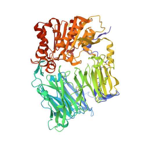
    3D4L

  • PubMed Abstract: 

    Probing with tool molecules, and by modeling and X-ray crystallography the binding modes of two structurally distinct series of DPP-4 inhibitors led to the discovery of a rare aromatic fluorine H-bond and the spatial requirement for better biaryl binding in the DPP-4 enzyme active site. These newly found binding elements were successfully incorporated into novel DPP-4 inhibitors.


  • Organizational Affiliation
    • gui-bai_liang@merck.comMerck Research Laboratories, Department of Medicinal Chemistry, Merck & Co., Inc., PO Box 2000, Rahway, NJ 07065, USA.

Macromolecule Content 

  • Total Structure Weight: 174.83 kDa 
  • Atom Count: 13,340 
  • Modeled Residue Count: 1,456 
  • Deposited Residue Count: 1,456 
  • Unique protein chains: 1

Macromolecules

Find similar proteins by:|  3D Structure
Entity ID: 1
MoleculeChains  Sequence LengthOrganismDetailsImage
Dipeptidyl peptidase 4 soluble form
A, B
728Homo sapiensMutation(s): 1 
Gene Names: DPP4ADCP2CD26
EC: 3.4.14.5
UniProt & NIH Common Fund Data Resources
Find proteins for P27487 (Homo sapiens)
Explore P27487 
Go to UniProtKB:  P27487
PHAROS:  P27487
GTEx:  ENSG00000197635 
Entity Groups
Sequence Clusters30% Identity50% Identity70% Identity90% Identity95% Identity100% Identity
UniProt GroupP27487
Glycosylation
Glycosylation Sites: 7Go to GlyGen: P27487-1
Sequence Annotations
Expand
Reference Sequence

Oligosaccharides

Help  
Entity ID: 2
MoleculeChains Length2D Diagram GlycosylationD Interactions
2-acetamido-2-deoxy-alpha-D-glucopyranose-(1-4)-2-acetamido-2-deoxy-beta-D-glucopyranose
C, F
2N-Glycosylation
Glycosylation Resources
GlyTouCan: G07375KG
GlyCosmos: G07375KG
GlyGen: G07375KG
Entity ID: 3
MoleculeChains Length2D Diagram GlycosylationD Interactions
2-acetamido-2-deoxy-beta-D-glucopyranose-(1-4)-2-acetamido-2-deoxy-beta-D-glucopyranose
D, E, G, H, I
D, E, G, H, I, J, K
2N-Glycosylation
Glycosylation Resources
GlyTouCan: G42666HT
GlyCosmos: G42666HT
GlyGen: G42666HT

Small Molecules

Ligands 3 Unique
IDChains Name / Formula / InChI Key2D Diagram3D Interactions
605

Query on 605


Download:Ideal Coordinates CCD File
P [auth A],
T [auth B]
4'-[(1R)-1-amino-2-(2,5-difluorophenyl)ethyl]biphenyl-3-carboxamide
C21 H18 F2 N2 O
JYKFWUXBFJJDTP-HXUWFJFHSA-N
NAG

Query on NAG


Download:Ideal Coordinates CCD File
L [auth A]
M [auth A]
N [auth A]
Q [auth B]
R [auth B]
L [auth A],
M [auth A],
N [auth A],
Q [auth B],
R [auth B],
S [auth B]
2-acetamido-2-deoxy-beta-D-glucopyranose
C8 H15 N O6
OVRNDRQMDRJTHS-FMDGEEDCSA-N
NA

Query on NA


Download:Ideal Coordinates CCD File
O [auth A]SODIUM ION
Na
FKNQFGJONOIPTF-UHFFFAOYSA-N

Experimental Data & Validation

Experimental Data

  • Method: X-RAY DIFFRACTION
  • Resolution: 2.00 Å
  • R-Value Free:  0.217 (Depositor), 0.214 (DCC) 
  • R-Value Work:  0.192 (Depositor), 0.190 (DCC) 
  • R-Value Observed: 0.220 (Depositor) 
Space Group: P 21 21 21
Unit Cell:
Length ( Å )Angle ( ˚ )
a = 118.435α = 90
b = 125.749β = 90
c = 137.175γ = 90
Software Package:
Software NamePurpose
MAR345dtbdata collection
CNXrefinement
HKL-2000data reduction
SCALEPACKdata scaling
CNXphasing

Structure Validation

View Full Validation Report


Ligand Structure Quality Assessment 


Entry History 

Deposition Data

  • Released Date: 2008-07-01 
  • Deposition Author(s): Scapin, G.

Revision History  (Full details and data files)

  • Version 1.0: 2008-07-01
    Type: Initial release
  • Version 1.1: 2011-07-13
    Changes: Non-polymer description, Version format compliance
  • Version 2.0: 2020-07-29
    Type: Remediation
    Reason: Carbohydrate remediation
    Changes: Advisory, Atomic model, Data collection, Derived calculations, Structure summary
  • Version 2.1: 2021-10-20
    Changes: Database references, Structure summary
  • Version 2.2: 2023-08-30
    Changes: Data collection, Refinement description
  • Version 2.3: 2024-10-30
    Changes: Structure summary