3D2D

Structure of berberine bridge enzyme in complex with (S)-reticuline


Experimental Data Snapshot

  • Method: X-RAY DIFFRACTION
  • Resolution: 2.80 Å
  • R-Value Free: 0.241 
  • R-Value Work: 0.188 
  • R-Value Observed: 0.191 

wwPDB Validation   3D Report Full Report


Ligand Structure Quality Assessment 


This is version 2.0 of the entry. See complete history


Literature

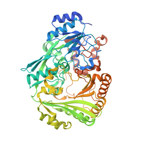
A concerted mechanism for berberine bridge enzyme

Winkler, A.Lyskowski, A.Riedl, S.Puhl, M.Kutchan, T.M.Macheroux, P.Gruber, K.

(2008) Nat Chem Biol 4: 739-741

  • DOI: https://doi.org/10.1038/nchembio.123
  • Primary Citation of Related Structures:  
    3D2D, 3D2H, 3D2J

  • PubMed Abstract: 

    Berberine bridge enzyme catalyzes the conversion of (S)-reticuline to (S)-scoulerine by formation of a carbon-carbon bond between the N-methyl group and the phenolic ring. We elucidated the structure of berberine bridge enzyme from Eschscholzia californica and determined the kinetic rates for three active site protein variants. Here we propose a catalytic mechanism combining base-catalyzed proton abstraction with concerted carbon-carbon coupling accompanied by hydride transfer from the N-methyl group to the N5 atom of the FAD cofactor.


  • Organizational Affiliation

    Institute of Biochemistry, Graz University of Technology, Petersgasse 12/II, 8010 Graz, Austria.


Macromolecules
Find similar proteins by:  (by identity cutoff)  |  3D Structure
Entity ID: 1
MoleculeChains Sequence LengthOrganismDetailsImage
berberine bridge-forming enzyme538Eschscholzia californicaMutation(s): 0 
Gene Names: BBE1
EC: 1.21.3.3
UniProt
Find proteins for P30986 (Eschscholzia californica)
Explore P30986 
Go to UniProtKB:  P30986
Entity Groups  
Sequence Clusters30% Identity50% Identity70% Identity90% Identity95% Identity100% Identity
UniProt GroupP30986
Sequence Annotations
Expand
  • Reference Sequence
Oligosaccharides

Help

Entity ID: 2
MoleculeChains Length2D Diagram Glycosylation3D Interactions
alpha-D-mannopyranose-(1-2)-alpha-D-mannopyranose-(1-2)-alpha-D-mannopyranose-(1-3)-alpha-D-mannopyranose-(1-4)-2-acetamido-2-deoxy-beta-D-glucopyranose-(1-4)-2-acetamido-2-deoxy-beta-D-glucopyranose
B
6N-Glycosylation
Glycosylation Resources
GlyTouCan:  G60005GQ
GlyCosmos:  G60005GQ
GlyGen:  G60005GQ
Small Molecules
Ligands 3 Unique
IDChains Name / Formula / InChI Key2D Diagram3D Interactions
FAD
Query on FAD

Download Ideal Coordinates CCD File 
D [auth A]FLAVIN-ADENINE DINUCLEOTIDE
C27 H33 N9 O15 P2
VWWQXMAJTJZDQX-UYBVJOGSSA-N
REN
Query on REN

Download Ideal Coordinates CCD File 
E [auth A](S)-reticuline
C19 H23 N O4
BHLYRWXGMIUIHG-HNNXBMFYSA-N
NAG
Query on NAG

Download Ideal Coordinates CCD File 
C [auth A]2-acetamido-2-deoxy-beta-D-glucopyranose
C8 H15 N O6
OVRNDRQMDRJTHS-FMDGEEDCSA-N
Experimental Data & Validation

Experimental Data

  • Method: X-RAY DIFFRACTION
  • Resolution: 2.80 Å
  • R-Value Free: 0.241 
  • R-Value Work: 0.188 
  • R-Value Observed: 0.191 
  • Space Group: C 1 2 1
Unit Cell:
Length ( Å )Angle ( ˚ )
a = 98.938α = 90
b = 93.649β = 100.62
c = 63.155γ = 90
Software Package:
Software NamePurpose
MAR345dtbdata collection
PHASERphasing
PHENIXrefinement
DENZOdata reduction
SCALEPACKdata scaling

Structure Validation

View Full Validation Report


Ligand Structure Quality Assessment 


Entry History 

Deposition Data

Revision History  (Full details and data files)

  • Version 1.0: 2008-10-28
    Type: Initial release
  • Version 1.1: 2011-07-13
    Changes: Non-polymer description, Version format compliance
  • Version 2.0: 2020-07-29
    Type: Remediation
    Reason: Carbohydrate remediation
    Changes: Advisory, Atomic model, Data collection, Database references, Derived calculations, Structure summary