3CV5 | pdb_00003cv5

GOLGI MANNOSIDASE II D204A catalytic nucleophile mutant complex with 3alpha,6alpha-mannopentaose


Experimental Data Snapshot

  • Method: X-RAY DIFFRACTION
  • Resolution: 1.60 Å
  • R-Value Free: 
    0.180 (Depositor), 0.163 (DCC) 
  • R-Value Work: 
    0.159 (Depositor), 0.159 (DCC) 
  • R-Value Observed: 
    0.159 (Depositor) 

Starting Model: experimental
View more details

wwPDB Validation 3D Report Full Report

Validation slider image for 3CV5

Ligand Structure Quality Assessment 


This is version 2.3 of the entry. See complete history

Literature

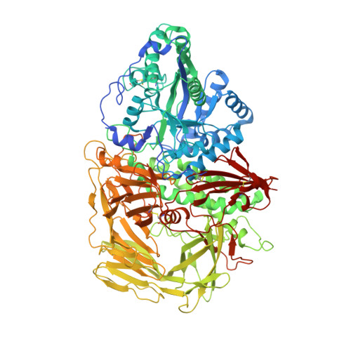
Golgi alpha-mannosidase II cleaves two sugars sequentially in the same catalytic site.

Shah, N.Kuntz, D.A.Rose, D.R.

(2008) Proc Natl Acad Sci U S A 105: 9570-9575

  • DOI: https://doi.org/10.1073/pnas.0802206105
  • Primary Citation Related Structures: 
    3CV5, 3CZN, 3CZS

  • PubMed Abstract: 

    Golgi alpha-mannosidase II (GMII) is a key glycosyl hydrolase in the N-linked glycosylation pathway. It catalyzes the removal of two different mannosyl linkages of GlcNAcMan(5)GlcNAc(2), which is the committed step in complex N-glycan synthesis. Inhibition of this enzyme has shown promise in certain cancers in both laboratory and clinical settings. Here we present the high-resolution crystal structure of a nucleophile mutant of Drosophila melanogaster GMII (dGMII) bound to its natural oligosaccharide substrate and an oligosaccharide precursor as well as the structure of the unliganded mutant. These structures allow us to identify three sugar-binding subsites within the larger active site cleft. Our results allow for the formulation of the complete catalytic process of dGMII, which involves a specific order of bond cleavage, and a major substrate rearrangement in the active site. This process is likely conserved for all GMII enzymes-but not in the structurally related lysosomal mannosidase-and will form the basis for the design of specific inhibitors against GMII.


  • Organizational Affiliation
    • Department of Medical Biophysics, University of Toronto, Toronto, ON, Canada.

Macromolecule Content 

  • Total Structure Weight: 120.89 kDa 
  • Atom Count: 9,644 
  • Modeled Residue Count: 1,015 
  • Deposited Residue Count: 1,045 
  • Unique protein chains: 1

Macromolecules

Find similar proteins by:|  3D Structure
Entity ID: 1
MoleculeChains  Sequence LengthOrganismDetailsImage
Alpha-mannosidase 21,045Drosophila melanogasterMutation(s): 1 
Gene Names: alpha-Man-IIGmII
EC: 3.2.1.114
UniProt
Find proteins for Q24451 (Drosophila melanogaster)
Explore Q24451 
Go to UniProtKB:  Q24451
Entity Groups
Sequence Clusters30% Identity50% Identity70% Identity90% Identity95% Identity100% Identity
UniProt GroupQ24451
Glycosylation
Glycosylation Sites: 1Go to GlyGen: Q24451-1
Sequence Annotations
Expand
Reference Sequence

Oligosaccharides

Help  
Entity ID: 2
MoleculeChains Length2D Diagram GlycosylationD Interactions
alpha-D-mannopyranose-(1-3)-[alpha-D-mannopyranose-(1-6)]alpha-D-mannopyranose-(1-6)-[alpha-D-mannopyranose-(1-3)]beta-D-mannopyranose
B
5N/A
Glycosylation Resources
GlyTouCan: G23046QV
GlyCosmos: G23046QV
GlyGen: G23046QV

Experimental Data & Validation

Experimental Data

  • Method: X-RAY DIFFRACTION
  • Resolution: 1.60 Å
  • R-Value Free:  0.180 (Depositor), 0.163 (DCC) 
  • R-Value Work:  0.159 (Depositor), 0.159 (DCC) 
  • R-Value Observed: 0.159 (Depositor) 
Space Group: P 21 21 21
Unit Cell:
Length ( Å )Angle ( ˚ )
a = 68.687α = 90
b = 109.958β = 90
c = 139.266γ = 90
Software Package:
Software NamePurpose
SAINTdata scaling
REFMACrefinement
PDB_EXTRACTdata extraction
ADSCdata collection
DENZOdata reduction
CNSphasing

Structure Validation

View Full Validation Report


Ligand Structure Quality Assessment 


Entry History 

Deposition Data

Revision History  (Full details and data files)

  • Version 1.0: 2008-06-24
    Type: Initial release
  • Version 1.1: 2011-07-13
    Changes: Non-polymer description, Version format compliance
  • Version 2.0: 2020-07-29
    Type: Remediation
    Reason: Carbohydrate remediation
    Changes: Advisory, Atomic model, Data collection, Database references, Derived calculations, Non-polymer description, Structure summary
  • Version 2.1: 2021-10-20
    Changes: Database references, Structure summary
  • Version 2.2: 2023-08-30
    Changes: Data collection, Refinement description
  • Version 2.3: 2024-10-30
    Changes: Structure summary