


Crystal structure of a Putative Branched-Chain Amino Acid Aminotransferase (TM0831) from Thermotoga maritima at 2.15 A resolution
Joint Center for Structural Genomics (JCSG)To be published.
Experimental Data Snapshot
Entity ID: 1 | |||||
|---|---|---|---|---|---|
| Molecule | Chains | Sequence Length | Organism | Details | Image |
| Putative branched-chain-amino-acid aminotransferase | 285 | Thermotoga maritima MSB8 | Mutation(s): 0 Gene Names: TM0831, ilvE EC: 2.6.1.42 | 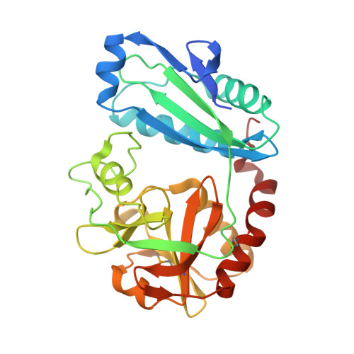 | |
UniProt | |||||
Entity Groups | |||||
| Sequence Clusters | 30% Identity50% Identity70% Identity90% Identity95% Identity100% Identity | ||||
| UniProt Group | P74921 | ||||
Sequence AnnotationsExpand | |||||
Reference Sequence | |||||
| Ligands 5 Unique | |||||
|---|---|---|---|---|---|
| ID | Chains | Name / Formula / InChI Key | 2D Diagram | 3D Interactions | |
| PLP Download:Ideal Coordinates CCD File | E [auth A], Q [auth C] | PYRIDOXAL-5'-PHOSPHATE C8 H10 N O6 P NGVDGCNFYWLIFO-UHFFFAOYSA-N |  | ||
| CIT Download:Ideal Coordinates CCD File | G [auth A], M [auth B], W [auth D] | CITRIC ACID C6 H8 O7 KRKNYBCHXYNGOX-UHFFFAOYSA-N |  | ||
| MPD Download:Ideal Coordinates CCD File | H [auth A] I [auth A] N [auth B] O [auth B] S [auth C] | (4S)-2-METHYL-2,4-PENTANEDIOL C6 H14 O2 SVTBMSDMJJWYQN-YFKPBYRVSA-N |  | ||
| CL Download:Ideal Coordinates CCD File | J [auth B], K [auth B], P [auth C], U [auth D] | CHLORIDE ION Cl VEXZGXHMUGYJMC-UHFFFAOYSA-M |  | ||
| UNL Download:Ideal Coordinates CCD File | F [auth A], L [auth B], R [auth C], V [auth D] | Unknown ligand VEXZGXHMUGYJMC-UHFFFAOYSA-M | |||
| Modified Residues 1 Unique | |||||
|---|---|---|---|---|---|
| ID | Chains | Type | Formula | 2D Diagram | Parent |
| MSE Query on MSE | A, B, C, D | L-PEPTIDE LINKING | C5 H11 N O2 Se |  | MET |
| Length ( Å ) | Angle ( ˚ ) |
|---|---|
| a = 107.71 | α = 90 |
| b = 129.76 | β = 90 |
| c = 298.69 | γ = 90 |
| Software Name | Purpose |
|---|---|
| REFMAC | refinement |
| PHENIX | refinement |
| SHELX | phasing |
| MolProbity | model building |
| XSCALE | data scaling |
| PDB_EXTRACT | data extraction |
| MAR345 | data collection |
| XDS | data reduction |
| SHARP | phasing |
| SHELXD | phasing |


