X-ray crystallographic and solution state nuclear magnetic resonance spectroscopic investigations of NADP+ binding to ferredoxin NADP reductase from Pseudomonas aeruginosa
Wang, A., Rodriguez, J.C., Han, H., Schonbrunn, E., Rivera, M.(2008) Biochemistry 47: 8080-8093
- PubMed: 18605699
- DOI: https://doi.org/10.1021/bi8007356
- Primary Citation of Related Structures:
3CRZ - PubMed Abstract:
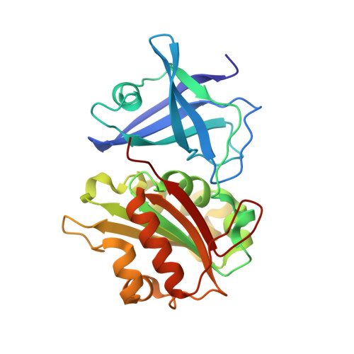
The ferredoxin nicotinamide adenine dinucleotide phosphate reductase from Pseudomonas aeruginosa ( pa-FPR) in complex with NADP (+) has been characterized by X-ray crystallography and in solution by NMR spectroscopy. The structure of the complex revealed that pa-FPR harbors a preformed NADP (+) binding pocket where the cofactor binds with minimal structural perturbation of the enzyme. These findings were complemented by obtaining sequential backbone resonance assignments of this 29518 kDa enzyme, which enabled the study of the pa-FPR-NADP complex by monitoring chemical shift perturbations induced by addition of NADP (+) or the inhibitor adenine dinucleotide phosphate (ADP) to pa-FPR. The results are consistent with a preformed NADP (+) binding site and also demonstrate that the pa-FPR-NADP complex is largely stabilized by interactions between the protein and the 2'-P AMP portion of the cofactor. Analysis of the crystal structure also shows a vast network of interactions between the two cofactors, FAD and NADP (+), and the characteristic AFVEK (258) C'-terminal extension that is typical of bacterial FPRs but is absent in their plastidic ferredoxin NADP (+) reductase (FNR) counterparts. The conformations of NADP (+) and FAD in pa-FPR place their respective nicotinamide and isoalloxazine rings 15 A apart and separated by residues in the C'-terminal extension. The network of interactions among NADP (+), FAD, and residues in the C'-terminal extension indicate that the gross conformational rearrangement that would be necessary to place the nicotinamide and isoalloxazine rings parallel and adjacent to one another for direct hydride transfer between NADPH and FAD in pa-FPR is highly unlikely. This conclusion is supported by observations made in the NMR spectra of pa-FPR and the pa-FPR-NADP complex, which strongly suggest that residues in the C'-terminal sequence do not undergo conformational exchange in the presence or absence of NADP (+). These findings are discussed in the context of a possible stepwise electron-proton-electron transfer of hydride in the oxidation of NADPH by FPR enzymes.
- Ralph N. Adams Institute for Bioanalytical Chemistry and Department of Chemistry, University of Kansas, Multidisciplinary Research Building, 2030 Becker Drive, Room 220 E, Lawrence, Kansas 66047, USA.
Organizational Affiliation:


