Structure of Escherichia coli tyrosine kinase Etk reveals a novel activation mechanism.
Lee, D.C., Zheng, J., She, Y.M., Jia, Z.(2008) EMBO J 27: 1758-1766
- PubMed: 18497741 Search on PubMedSearch on PubMed Central
- DOI: https://doi.org/10.1038/emboj.2008.97
- Primary Citation Related Structures:
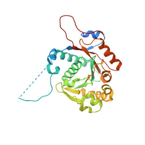
3CIO - PubMed Abstract:
While protein tyrosine (Tyr) kinases (PTKs) have been extensively characterized in eukaryotes, far less is known about their emerging counterparts in prokaryotes. The inner-membrane Wzc/Etk protein belongs to the bacterial PTK family, which has an important function in regulating the polymerization and transport of virulence-determining capsular polysaccharide (CPS). The kinase uses a unique two-step activation process involving intra-phosphorylation of a Tyr residue, although the molecular mechanism remains unknown. Herein, we report the first crystal structure of a bacterial PTK, the C-terminal kinase domain of Escherichia coli Tyr kinase (Etk) at 2.5-A resolution. The fold of the Etk kinase domain differs markedly from that of eukaryotic PTKs. Based on the observed structure and supporting mass spectrometric evidence of Etk, a unique activation mechanism is proposed that involves the phosphorylated Tyr residue, Y574, at the active site and its specific interaction with a previously unidentified key Arg residue, R614, to unblock the active site. Both in vitro kinase activity and in vivo antibiotics resistance studies using structure-guided mutants further support the novel activation mechanism.
- Department of Biochemistry, Queen's University, Kingston, Ontario, Canada.
Organizational Affiliation: