


Crystal structure of glycerophosphoryl diester phosphodiesterase (YP_677622.1) from Cytophaga hutchinsonii ATCC 33406 at 1.50 A resolution
Joint Center for Structural Genomics (JCSG)To be published.
Experimental Data Snapshot
wwPDB Validation 3D Report Full Report

Entity ID: 1 | |||||
|---|---|---|---|---|---|
| Molecule | Chains | Sequence Length | Organism | Details | Image |
| Glycerophosphodiester phosphodiesterase | 272 | Cytophaga hutchinsonii ATCC 33406 | Mutation(s): 0 Gene Names: YP_677622.1, ugpQ, CHU_1005 EC: 3.1.4.46 | 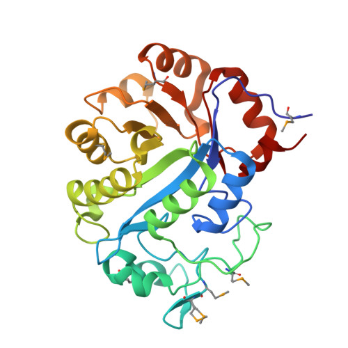 | |
| Ligands 3 Unique | |||||
|---|---|---|---|---|---|
| ID | Chains | Name / Formula / InChI Key | 2D Diagram | 3D Interactions | |
| CIT Download:Ideal Coordinates CCD File | B [auth A] | CITRIC ACID C6 H8 O7 KRKNYBCHXYNGOX-UHFFFAOYSA-N |  | ||
| GOL Download:Ideal Coordinates CCD File | S [auth A] | GLYCEROL C3 H8 O3 PEDCQBHIVMGVHV-UHFFFAOYSA-N |  | ||
| EDO Download:Ideal Coordinates CCD File | C [auth A] D [auth A] E [auth A] F [auth A] G [auth A] | 1,2-ETHANEDIOL C2 H6 O2 LYCAIKOWRPUZTN-UHFFFAOYSA-N |  | ||
| Modified Residues 1 Unique | |||||
|---|---|---|---|---|---|
| ID | Chains | Type | Formula | 2D Diagram | Parent |
| MSE Query on MSE | A | L-PEPTIDE LINKING | C5 H11 N O2 Se |  | MET |
| Length ( Å ) | Angle ( ˚ ) |
|---|---|
| a = 49.532 | α = 90 |
| b = 61.211 | β = 90 |
| c = 113.699 | γ = 90 |
| Software Name | Purpose |
|---|---|
| REFMAC | refinement |
| PHENIX | refinement |
| SOLVE | phasing |
| MolProbity | model building |
| SCALA | data scaling |
| PDB_EXTRACT | data extraction |
| MAR345 | data collection |
| MOSFLM | data reduction |


