3CAE | pdb_00003cae

Structure of NNQQNY as an insert in T7 endonuclease I


Experimental Data Snapshot

  • Method: X-RAY DIFFRACTION
  • Resolution: 3.00 Å
  • R-Value Free: 
    0.284 (Depositor), 0.272 (DCC) 
  • R-Value Work: 
    0.240 (Depositor), 0.233 (DCC) 

Starting Model: experimental
View more details

wwPDB Validation 3D Report Full Report

Validation slider image for 3CAE

This is version 1.4 of the entry. See complete history

Literature

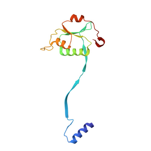
The structure of a fibril-forming sequence, NNQQNY, in the context of a globular fold.

Guo, Z.Eisenberg, D.

(2008) Protein Sci 17: 1617-1623

  • DOI: https://doi.org/10.1110/ps.036368.108
  • Primary Citation Related Structures: 
    3CAE

  • PubMed Abstract: 

    Numerous human disorders are associated with the formation of protein fibrils. The fibril-forming capacity of a protein has been found in recent studies to be determined by a short segment of residues that forms a dual beta-sheet, called a steric zipper, in the spine of the fibril. The question arises as to whether a fibril-forming segment, when inserted within the sequence of a globular protein, will invariably cause the protein to form fibrils. Here we investigate this question by inserting the known fibril-forming segment NNQQNY into the globular enzyme, T7 endonuclease I. From earlier studies, we know that in its fibril form, NNQQNY is in an extended conformation. We first found that the inserted NNQQNY stimulates fibril formation of T7 endonuclease I in solution. Thus NNQQNY within T7 endonuclease I can exist in an extended conformation, capable of forming the steric zipper in the core of a fibril. We also found that T7 endonuclease I folds into a decamer that does not form fibrils. We determined the structure of the decamer by X-ray crystallography, finding an unusual oligomer without point group symmetry, and finding that the NNQQNY segments within the decamer adopt two twisted conformations, neither is apparently able to fibrillize. We conclude that twisting of fibril forming sequences from the fully extended conformation, imposed by the context of their placement in proteins, can interfere with fibril formation.


  • Organizational Affiliation
    • Howard Hughes Medical Institute, UCLA-DOE Institute for Genomics and Proteomics, Molecular Biology Institute, UCLA, Los Angeles, California 90095-1570, USA.

Macromolecule Content 

  • Total Structure Weight: 156.51 kDa 
  • Atom Count: 11,075 
  • Modeled Residue Count: 1,320 
  • Deposited Residue Count: 1,320 
  • Unique protein chains: 1

Macromolecules

Find similar proteins by:|  3D Structure
Entity ID: 1
MoleculeChains  Sequence LengthOrganismDetailsImage
Endonuclease I
A, B, C, D, E
A, B, C, D, E, F, G, H, I, J
132Escherichia phage T7Mutation(s): 0 
EC: 3.1.21.2
UniProt
Find proteins for P00641 (Escherichia phage T7)
Explore P00641 
Go to UniProtKB:  P00641
Entity Groups
Sequence Clusters30% Identity50% Identity70% Identity90% Identity95% Identity100% Identity
UniProt GroupP00641
Sequence Annotations
Expand
Reference Sequence

Experimental Data & Validation

Experimental Data

  • Method: X-RAY DIFFRACTION
  • Resolution: 3.00 Å
  • R-Value Free:  0.284 (Depositor), 0.272 (DCC) 
  • R-Value Work:  0.240 (Depositor), 0.233 (DCC) 
Space Group: P 1 21 1
Unit Cell:
Length ( Å )Angle ( ˚ )
a = 88.408α = 90
b = 140.343β = 93.37
c = 88.508γ = 90
Software Package:
Software NamePurpose
DENZOdata reduction
SCALEPACKdata scaling
CNSrefinement
PDB_EXTRACTdata extraction
BOSdata collection
PHASERphasing

Structure Validation

View Full Validation Report


Entry History 

Deposition Data

Revision History  (Full details and data files)

  • Version 1.0: 2008-07-22
    Type: Initial release
  • Version 1.1: 2011-07-13
    Changes: Version format compliance
  • Version 1.2: 2017-06-07
    Changes: Database references, Source and taxonomy, Structure summary
  • Version 1.3: 2017-10-25
    Changes: Refinement description
  • Version 1.4: 2023-08-30
    Changes: Data collection, Database references, Refinement description