3BZC

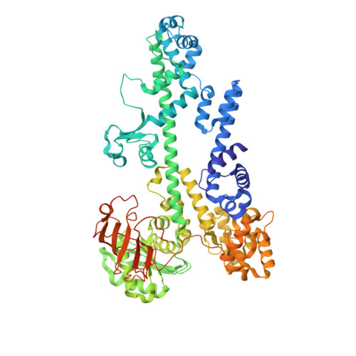
Crystal Structure of the Tex protein from Pseudomonas aeruginosa, crystal form I


Experimental Data Snapshot

  • Method: X-RAY DIFFRACTION
  • Resolution: 2.27 Å
  • R-Value Free: 0.274 
  • R-Value Work: 0.240 
  • R-Value Observed: 0.288 

wwPDB Validation   3D Report Full Report


This is version 1.3 of the entry. See complete history


Literature

Crystal structure and RNA binding of the Tex protein from Pseudomonas aeruginosa.

Johnson, S.J.Close, D.Robinson, H.Vallet-Gely, I.Dove, S.L.Hill, C.P.

(2008) J Mol Biol 377: 1460-1473

  • DOI: https://doi.org/10.1016/j.jmb.2008.01.096
  • Primary Citation of Related Structures:  
    3BZC, 3BZK

  • PubMed Abstract: 

    Tex is a highly conserved bacterial protein that likely functions in a variety of transcriptional processes. Here, we describe two crystal structures of the 86-kDa Tex protein from Pseudomonas aeruginosa at 2.3 and 2.5 A resolution, respectively. These structures reveal a relatively flat and elongated protein, with several potential nucleic acid binding motifs clustered at one end, including an S1 domain near the C-terminus that displays considerable structural flexibility. Tex binds nucleic acids, with a preference for single-stranded RNA, and the Tex S1 domain is required for this binding activity. Point mutants further demonstrate that the primary nucleic acid binding site corresponds to a surface of the S1 domain. Sequence alignment and modeling indicate that the eukaryotic Spt6 transcription factor adopts a similar core structure. Structural analysis further suggests that the RNA polymerase and nucleosome interacting regions of Spt6 flank opposite sides of the Tex-like scaffold. Therefore, the Tex structure may represent a conserved scaffold that binds single-stranded RNA to regulate transcription in both eukaryotic and prokaryotic organisms.


  • Organizational Affiliation

    Department of Chemistry and Biochemistry, Utah State University, Logan, UT 84322-0300, USA.


Macromolecules
Find similar proteins by:  (by identity cutoff)  |  3D Structure
Entity ID: 1
MoleculeChains Sequence LengthOrganismDetailsImage
Tex785Pseudomonas aeruginosaMutation(s): 0 
UniProt
Find proteins for Q9HTY8 (Pseudomonas aeruginosa (strain ATCC 15692 / DSM 22644 / CIP 104116 / JCM 14847 / LMG 12228 / 1C / PRS 101 / PAO1))
Explore Q9HTY8 
Go to UniProtKB:  Q9HTY8
Entity Groups  
Sequence Clusters30% Identity50% Identity70% Identity90% Identity95% Identity100% Identity
UniProt GroupQ9HTY8
Sequence Annotations
Expand
  • Reference Sequence
Experimental Data & Validation

Experimental Data

  • Method: X-RAY DIFFRACTION
  • Resolution: 2.27 Å
  • R-Value Free: 0.274 
  • R-Value Work: 0.240 
  • R-Value Observed: 0.288 
  • Space Group: P 21 21 21
Unit Cell:
Length ( Å )Angle ( ˚ )
a = 57.165α = 90
b = 131.849β = 90
c = 144.017γ = 90
Software Package:
Software NamePurpose
DENZOdata reduction
SCALEPACKdata scaling
PHENIXrefinement
PDB_EXTRACTdata extraction
ADSCdata collection
HKL-2000data reduction
SOLVEphasing

Structure Validation

View Full Validation Report


Entry History 

Deposition Data

Revision History  (Full details and data files)

  • Version 1.0: 2008-04-08
    Type: Initial release
  • Version 1.1: 2011-07-13
    Changes: Version format compliance
  • Version 1.2: 2017-10-25
    Changes: Refinement description
  • Version 1.3: 2024-02-21
    Changes: Data collection, Database references