Assessing energetic contributions to binding from a disordered region in a protein-protein interaction
Cho, S., Swaminathan, C.P., Bonsor, D.A., Kerzic, M.C., Guan, R., Yang, J., Kieke, M.C., Andersen, P.S., Kranz, D.M., Mariuzza, R.A., Sundberg, E.J.(2010) Biochemistry 49: 9256-9268
- PubMed: 20836565 Search on PubMedSearch on PubMed Central
- DOI: https://doi.org/10.1021/bi1008968
- Primary Citation Related Structures:
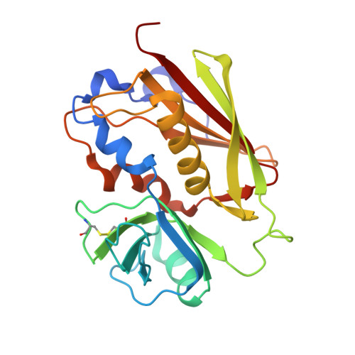
3BVG - PubMed Abstract:
Many functional proteins are at least partially disordered prior to binding. Although the structural transitions upon binding of disordered protein regions can influence the affinity and specificity of protein complexes, their precise energetic contributions to binding are unknown. Here, we use a model protein-protein interaction system in which a locally disordered region has been modified by directed evolution to quantitatively assess the thermodynamic and structural contributions to binding of disorder-to-order transitions. Through X-ray structure determination of the protein binding partners before and after complex formation and isothermal titration calorimetry of the interactions, we observe a correlation between protein ordering and binding affinity for complexes along this affinity maturation pathway. Additionally, we show that discrepancies between observed and calculated heat capacities based on buried surface area changes in the protein complexes can be explained largely by heat capacity changes that would result solely from folding the locally disordered region. Previously developed algorithms for predicting binding energies of protein-protein interactions, however, are unable to correctly model the energetic contributions of the structural transitions in our model system. While this highlights the shortcomings of current computational methods in modeling conformational flexibility, it suggests that the experimental methods used here could provide training sets of molecular interactions for improving these algorithms and further rationalizing molecular recognition in protein-protein interactions.
- W. M. Keck Laboratory for Structural Biology, University of Maryland Institute for Bioscience and Biotechnology Research, Rockville, MD 20850, USA.
Organizational Affiliation: