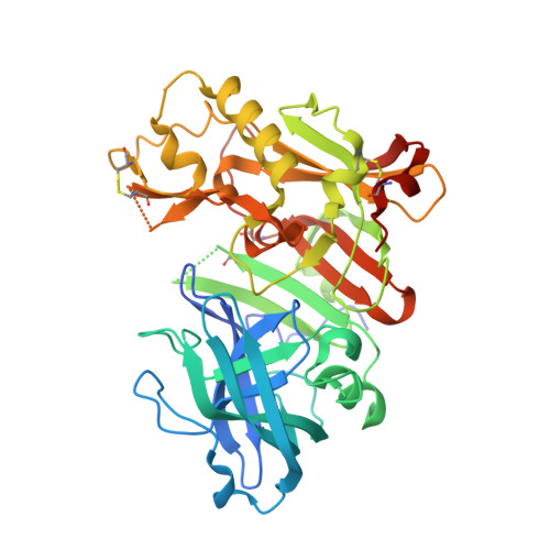
3BUH

BACE-1 complexed with compound 4


Experimental Data Snapshot

  • Method: X-RAY DIFFRACTION
  • Resolution: 2.30 Å
  • R-Value Free: 0.238 
  • R-Value Work: 0.199 
  • R-Value Observed: 0.201 

wwPDB Validation   3D Report Full Report


This is version 1.2 of the entry. See complete history


Literature

Tyramine fragment binding to BACE-1

Kuglstatter, A.Stahl, M.Peters, J.U.Huber, W.Stihle, M.Schlatter, D.Benz, J.Ruf, A.Roth, D.Enderle, T.Hennig, M.

(2008) Bioorg Med Chem Lett 18: 1304-1307

  • DOI: https://doi.org/10.1016/j.bmcl.2008.01.032
  • Primary Citation of Related Structures:  
    3BRA, 3BUF, 3BUG, 3BUH

  • PubMed Abstract: 
  • Fragment screening revealed that tyramine binds to the active site of the Alzheimer's disease drug target BACE-1. Hit expansion by selection of compounds from the Roche compound library identified tyramine derivatives with improved binding affinities as monitored by surface plasmon resonance ...

    Fragment screening revealed that tyramine binds to the active site of the Alzheimer's disease drug target BACE-1. Hit expansion by selection of compounds from the Roche compound library identified tyramine derivatives with improved binding affinities as monitored by surface plasmon resonance. X-ray structures show that the amine of the tyramine fragment hydrogen-bonds to the catalytic water molecule. Structure-guided ligand design led to the synthesis of further low molecular weight compounds that are starting points for chemical leads.


    Organizational Affiliation

    F. Hoffmann-La Roche, Pharma Research Basel, Grenzacherstr., 4070 Basel, Switzerland.


Macromolecules
Find similar proteins by:  (by identity cutoff)  |  3D Structure
Entity ID: 1
MoleculeChainsSequence LengthOrganismDetailsImage
beta-secretase 1409Homo sapiensMutation(s): 1 
Gene Names: BACE1BACEKIAA1149
EC: 3.4.23.46
UniProt & NIH Common Fund Data Resources
Find proteins for P56817 (Homo sapiens)
Explore P56817 
Go to UniProtKB:  P56817
PHAROS:  P56817
Entity Groups  
Sequence Clusters30% Identity50% Identity70% Identity90% Identity95% Identity100% Identity
UniProt GroupP56817
Protein Feature View
Expand
  • Reference Sequence
Small Molecules
Ligands 1 Unique
IDChainsName / Formula / InChI Key2D Diagram3D Interactions
AED
Query on AED

Download Ideal Coordinates CCD File 
B [auth A]4-(2-aminoethyl)-2-cyclohexylphenol
C14 H21 N O
DOCCSEDGBYCYLS-UHFFFAOYSA-N
 Ligand Interaction
Binding Affinity Annotations 
IDSourceBinding Affinity
AED PDBBind:  3BUH Kd: 2.20e+5 (nM) from 1 assay(s)
BindingDB:  3BUH Kd: 2.20e+5 (nM) from 1 assay(s)
Binding MOAD:  3BUH Kd: 2.20e+5 (nM) from 1 assay(s)
Experimental Data & Validation

Experimental Data

  • Method: X-RAY DIFFRACTION
  • Resolution: 2.30 Å
  • R-Value Free: 0.238 
  • R-Value Work: 0.199 
  • R-Value Observed: 0.201 
  • Space Group: P 61 2 2
Unit Cell:
Length ( Å )Angle ( ˚ )
a = 102.491α = 90
b = 102.491β = 90
c = 170.063γ = 120
Software Package:
Software NamePurpose
REFMACrefinement
DENZOdata reduction
SCALEPACKdata scaling

Structure Validation

View Full Validation Report


Entry History 

Deposition Data

Revision History  (Full details and data files)

  • Version 1.0: 2008-03-11
    Type: Initial release
  • Version 1.1: 2011-07-13
    Changes: Version format compliance
  • Version 1.2: 2021-11-10
    Changes: Database references, Derived calculations