3BUH | pdb_00003buh

BACE-1 complexed with compound 4


Experimental Data Snapshot

  • Method: X-RAY DIFFRACTION
  • Resolution: 2.30 Å
  • R-Value Free: 
    0.238 (Depositor) 
  • R-Value Work: 
    0.199 (Depositor) 
  • R-Value Observed: 
    0.201 (Depositor) 

wwPDB Validation 3D Report Full Report

Validation slider image for 3BUH

This is version 1.3 of the entry. See complete history

Literature

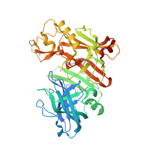
Tyramine fragment binding to BACE-1

Kuglstatter, A.Stahl, M.Peters, J.U.Huber, W.Stihle, M.Schlatter, D.Benz, J.Ruf, A.Roth, D.Enderle, T.Hennig, M.

(2008) Bioorg Med Chem Lett 18: 1304-1307

  • DOI: https://doi.org/10.1016/j.bmcl.2008.01.032
  • Primary Citation Related Structures: 
    3BRA, 3BUF, 3BUG, 3BUH

  • PubMed Abstract: 

    Fragment screening revealed that tyramine binds to the active site of the Alzheimer's disease drug target BACE-1. Hit expansion by selection of compounds from the Roche compound library identified tyramine derivatives with improved binding affinities as monitored by surface plasmon resonance. X-ray structures show that the amine of the tyramine fragment hydrogen-bonds to the catalytic water molecule. Structure-guided ligand design led to the synthesis of further low molecular weight compounds that are starting points for chemical leads.


  • Organizational Affiliation
    • F. Hoffmann-La Roche, Pharma Research Basel, Grenzacherstr., 4070 Basel, Switzerland.

Macromolecule Content 

  • Total Structure Weight: 45.77 kDa 
  • Atom Count: 3,103 
  • Modeled Residue Count: 370 
  • Deposited Residue Count: 409 
  • Unique protein chains: 1

Macromolecules

Find similar proteins by:|  3D Structure
Entity ID: 1
MoleculeChains  Sequence LengthOrganismDetailsImage
beta-secretase 1409Homo sapiensMutation(s): 1 
EC: 3.4.23.46
UniProt & NIH Common Fund Data Resources
Find proteins for P56817 (Homo sapiens)
Explore P56817 
Go to UniProtKB:  P56817
PHAROS:  P56817
GTEx:  ENSG00000186318 
Entity Groups
Sequence Clusters30% Identity50% Identity70% Identity90% Identity95% Identity100% Identity
UniProt GroupP56817
Sequence Annotations
Expand
Reference Sequence

Small Molecules

Ligands 1 Unique
IDChains Name / Formula / InChI Key2D Diagram3D Interactions
AED

Query on AED


Download:Ideal Coordinates CCD File
B [auth A]4-(2-aminoethyl)-2-cyclohexylphenol
C14 H21 N O
DOCCSEDGBYCYLS-UHFFFAOYSA-N
Binding Affinity Annotations 
IDSourceBinding Affinity
AED BindingDB:  3BUH Kd: 2.20e+5 (nM) from 1 assay(s)

Experimental Data & Validation

Experimental Data

  • Method: X-RAY DIFFRACTION
  • Resolution: 2.30 Å
  • R-Value Free:  0.238 (Depositor) 
  • R-Value Work:  0.199 (Depositor) 
  • R-Value Observed: 0.201 (Depositor) 
Space Group: P 61 2 2
Unit Cell:
Length ( Å )Angle ( ˚ )
a = 102.491α = 90
b = 102.491β = 90
c = 170.063γ = 120
Software Package:
Software NamePurpose
REFMACrefinement
DENZOdata reduction
SCALEPACKdata scaling

Structure Validation

View Full Validation Report


Entry History 

Deposition Data

Revision History  (Full details and data files)

  • Version 1.0: 2008-03-11
    Type: Initial release
  • Version 1.1: 2011-07-13
    Changes: Version format compliance
  • Version 1.2: 2021-11-10
    Changes: Database references, Derived calculations
  • Version 1.3: 2024-10-30
    Changes: Data collection, Structure summary