3BKM

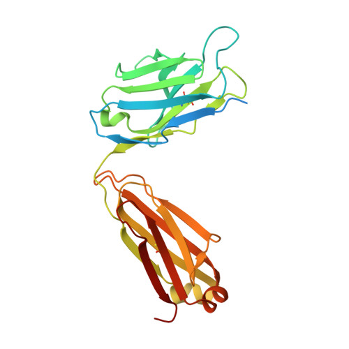
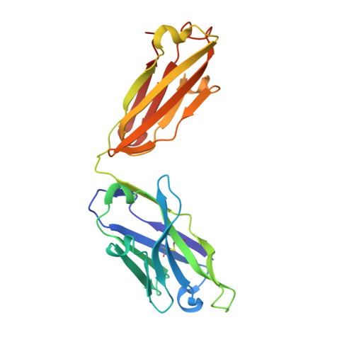
Structure of anti-amyloid-beta Fab WO2 (Form A, P212121)


Experimental Data Snapshot

  • Method: X-RAY DIFFRACTION
  • Resolution: 1.60 Å
  • R-Value Free: 0.226 
  • R-Value Work: 0.203 
  • R-Value Observed: 0.205 

Starting Model: experimental
View more details

wwPDB Validation   3D Report Full Report


This is version 1.2 of the entry. See complete history


Literature

Amyloid-beta-anti-amyloid-beta complex structure reveals an extended conformation in the immunodominant B-cell epitope.

Miles, L.A.Wun, K.S.Crespi, G.A.Fodero-Tavoletti, M.T.Galatis, D.Bagley, C.J.Beyreuther, K.Masters, C.L.Cappai, R.McKinstry, W.J.Barnham, K.J.Parker, M.W.

(2008) J Mol Biol 377: 181-192

  • DOI: https://doi.org/10.1016/j.jmb.2007.12.036
  • Primary Citation of Related Structures:  
    3BAE, 3BKC, 3BKJ, 3BKM

  • PubMed Abstract: 

    Alzheimer's disease (AD) is the most common form of dementia. Amyloid-beta (A beta) peptide, generated by proteolytic cleavage of the amyloid precursor protein, is central to AD pathogenesis. Most pharmaceutical activity in AD research has focused on A beta, its generation and clearance from the brain. In particular, there is much interest in immunotherapy approaches with a number of anti-A beta antibodies in clinical trials. We have developed a monoclonal antibody, called WO2, which recognises the A beta peptide. To this end, we have determined the three-dimensional structure, to near atomic resolution, of both the antibody and the complex with its antigen, the A beta peptide. The structures reveal the molecular basis for WO2 recognition and binding of A beta. The A beta peptide adopts an extended, coil-like conformation across its major immunodominant B-cell epitope between residues 2 and 8. We have also studied the antibody-bound A beta peptide in the presence of metals known to affect its aggregation state and show that WO2 inhibits these interactions. Thus, antibodies that target the N-terminal region of A beta, such as WO2, hold promise for therapeutic development.


  • Organizational Affiliation

    Biota Structural Biology Laboratory, St. Vincent's Institute of Medical Research, 9 Princes Street, Fitzroy, Victoria 3065, Australia.


Macromolecules
Find similar proteins by:  (by identity cutoff)  |  3D Structure
Entity ID: 1
MoleculeChains Sequence LengthOrganismDetailsImage
WO2 IgG2a Fab fragment Light Chain KappaA [auth L]252Mus musculusMutation(s): 0 
UniProt
Find proteins for A2NHM3 (Mus musculus)
Explore A2NHM3 
Go to UniProtKB:  A2NHM3
Entity Groups  
Sequence Clusters30% Identity50% Identity70% Identity90% Identity95% Identity100% Identity
UniProt GroupA2NHM3
Sequence Annotations
Expand
  • Reference Sequence
Find similar proteins by:  (by identity cutoff)  |  3D Structure
Entity ID: 2
MoleculeChains Sequence LengthOrganismDetailsImage
WO2 IgG2a Fab fragment Heavy ChainB [auth H]224Mus musculusMutation(s): 0 
UniProt
Find proteins for P84751 (Mus musculus)
Explore P84751 
Go to UniProtKB:  P84751
Entity Groups  
Sequence Clusters30% Identity50% Identity70% Identity90% Identity95% Identity100% Identity
UniProt GroupP84751
Sequence Annotations
Expand
  • Reference Sequence
Experimental Data & Validation

Experimental Data

  • Method: X-RAY DIFFRACTION
  • Resolution: 1.60 Å
  • R-Value Free: 0.226 
  • R-Value Work: 0.203 
  • R-Value Observed: 0.205 
  • Space Group: P 21 21 21
Unit Cell:
Length ( Å )Angle ( ˚ )
a = 52.447α = 90
b = 90.496β = 90
c = 123.279γ = 90
Software Package:
Software NamePurpose
REFMACrefinement
PDB_EXTRACTdata extraction
CrystalCleardata collection
DENZOdata reduction
SCALEPACKdata scaling
AMoREphasing

Structure Validation

View Full Validation Report


Entry History 

Revision History  (Full details and data files)

  • Version 1.0: 2008-04-15
    Type: Initial release
  • Version 1.1: 2011-07-13
    Changes: Version format compliance
  • Version 1.2: 2023-11-01
    Changes: Data collection, Database references, Derived calculations, Refinement description