3BIW

Crystal structure of the Neuroligin-1/Neurexin-1beta synaptic adhesion complex


Experimental Data Snapshot

  • Method: X-RAY DIFFRACTION
  • Resolution: 3.50 Å
  • R-Value Free: 0.276 
  • R-Value Work: 0.246 

wwPDB Validation   3D Report Full Report


Ligand Structure Quality Assessment 


This is version 2.0 of the entry. See complete history


Literature

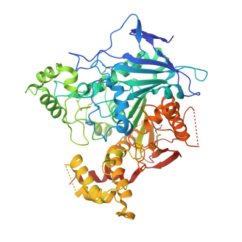
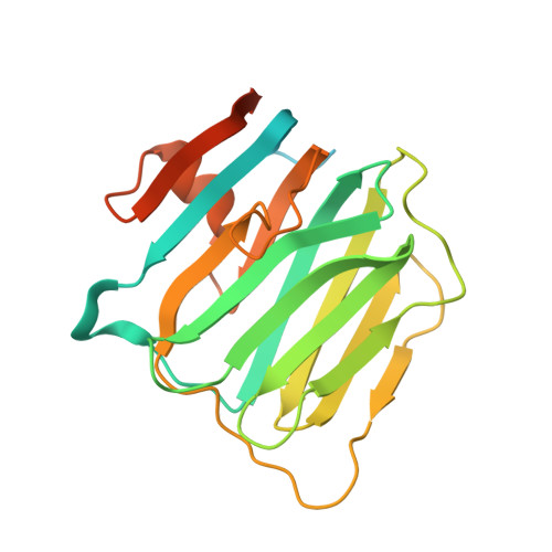
Structures of Neuroligin-1 and the Neuroligin-1/Neurexin-1beta Complex Reveal Specific Protein-Protein and Protein-Ca(2+) Interactions.

Arac, D.Boucard, A.A.Ozkan, E.Strop, P.Newell, E.Sudhof, T.C.Brunger, A.T.

(2007) Neuron 56: 992-1003

  • DOI: https://doi.org/10.1016/j.neuron.2007.12.002
  • Primary Citation of Related Structures:  
    3BIW, 3BIX

  • PubMed Abstract: 

    Neurexins and neuroligins provide trans-synaptic connectivity by the Ca2+-dependent interaction of their alternatively spliced extracellular domains. Neuroligins specify synapses in an activity-dependent manner, presumably by binding to neurexins. Here, we present the crystal structures of neuroligin-1 in isolation and in complex with neurexin-1 beta. Neuroligin-1 forms a constitutive dimer, and two neurexin-1 beta monomers bind to two identical surfaces on the opposite faces of the neuroligin-1 dimer to form a heterotetramer. The neuroligin-1/neurexin-1 beta complex exhibits a nanomolar affinity and includes a large binding interface that contains bound Ca2+. Alternatively spliced sites in neurexin-1 beta and in neuroligin-1 are positioned nearby the binding interface, explaining how they regulate the interaction. Structure-based mutations of neuroligin-1 at the interface disrupt binding to neurexin-1 beta, but not the folding of neuroligin-1 and confirm the validity of the binding interface of the neuroligin-1/neurexin-1 beta complex. Our results provide molecular insights for understanding the role of cell-adhesion proteins in synapse function.


  • Organizational Affiliation

    Howard Hughes Medical Institute, Stanford University, Stanford, CA 94305, USA.


Macromolecules
Find similar proteins by:  (by identity cutoff)  |  3D Structure
Entity ID: 1
MoleculeChains Sequence LengthOrganismDetailsImage
Neuroligin-1
A, B, C, D
574Rattus norvegicusMutation(s): 0 
Gene Names: Nlgn1
Membrane Entity: Yes 
UniProt
Find proteins for Q62765 (Rattus norvegicus)
Explore Q62765 
Go to UniProtKB:  Q62765
Entity Groups  
Sequence Clusters30% Identity50% Identity70% Identity90% Identity95% Identity100% Identity
UniProt GroupQ62765
Sequence Annotations
Expand
  • Reference Sequence
Find similar proteins by:  (by identity cutoff)  |  3D Structure
Entity ID: 2
MoleculeChains Sequence LengthOrganismDetailsImage
Neurexin-1-beta
E, F, G, H
243Rattus norvegicusMutation(s): 0 
Gene Names: Nrxn1
Membrane Entity: Yes 
UniProt
Find proteins for Q63372 (Rattus norvegicus)
Explore Q63372 
Go to UniProtKB:  Q63372
Entity Groups  
Sequence Clusters30% Identity50% Identity70% Identity90% Identity95% Identity100% Identity
UniProt GroupQ63372
Sequence Annotations
Expand
  • Reference Sequence
Oligosaccharides

Help

Entity ID: 3
MoleculeChains Length2D Diagram Glycosylation3D Interactions
2-acetamido-2-deoxy-beta-D-glucopyranose-(1-4)-2-acetamido-2-deoxy-beta-D-glucopyranose
I, J, K
2N-Glycosylation
Glycosylation Resources
GlyTouCan:  G42666HT
GlyCosmos:  G42666HT
GlyGen:  G42666HT
Small Molecules
Ligands 2 Unique
IDChains Name / Formula / InChI Key2D Diagram3D Interactions
NAG
Query on NAG

Download Ideal Coordinates CCD File 
L [auth A]
M [auth A]
N [auth A]
O [auth B]
P [auth B]
L [auth A],
M [auth A],
N [auth A],
O [auth B],
P [auth B],
Q [auth B],
R [auth C],
S [auth C],
T [auth C],
U [auth D],
V [auth D],
W [auth D]
2-acetamido-2-deoxy-beta-D-glucopyranose
C8 H15 N O6
OVRNDRQMDRJTHS-FMDGEEDCSA-N
CA
Query on CA

Download Ideal Coordinates CCD File 
AA [auth F]
BA [auth G]
CA [auth G]
X [auth E]
Y [auth E]
AA [auth F],
BA [auth G],
CA [auth G],
X [auth E],
Y [auth E],
Z [auth F]
CALCIUM ION
Ca
BHPQYMZQTOCNFJ-UHFFFAOYSA-N
Experimental Data & Validation

Experimental Data

  • Method: X-RAY DIFFRACTION
  • Resolution: 3.50 Å
  • R-Value Free: 0.276 
  • R-Value Work: 0.246 
  • Space Group: C 1 2 1
Unit Cell:
Length ( Å )Angle ( ˚ )
a = 229.83α = 90
b = 148.796β = 90.38
c = 123.602γ = 90
Software Package:
Software NamePurpose
DENZOdata reduction
SCALEPACKdata scaling
PHASERphasing
CNSrefinement
PDB_EXTRACTdata extraction
HKL-2000data reduction
HKL-2000data scaling

Structure Validation

View Full Validation Report


Ligand Structure Quality Assessment 


Entry History 

Deposition Data

Revision History  (Full details and data files)

  • Version 1.0: 2007-12-18
    Type: Initial release
  • Version 1.1: 2011-07-13
    Changes: Non-polymer description, Version format compliance
  • Version 1.2: 2017-08-23
    Changes: Refinement description, Source and taxonomy
  • Version 1.3: 2017-10-25
    Changes: Refinement description
  • Version 2.0: 2020-07-29
    Type: Remediation
    Reason: Carbohydrate remediation
    Changes: Atomic model, Data collection, Database references, Derived calculations, Structure summary