High-resolution structure of ybfF from Escherichia coli K12: a unique substrate-binding crevice generated by domain arrangement
Park, S.Y., Lee, S.H., Lee, J., Nishi, K., Kim, Y.S., Jung, C.H., Kim, J.S.(2008) J Mol Biol 376: 1426-1437
- PubMed: 18215690 Search on PubMed
- DOI: https://doi.org/10.1016/j.jmb.2007.12.062
- Primary Citation Related Structures:
3BF7, 3BF8 - PubMed Abstract:
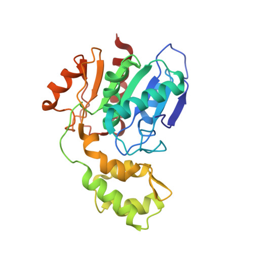
Esterases are one of the most common enzymes and are involved in diverse cellular functions. ybfF protein from Escherichia coli (Ec_ybfF) belongs to the esterase family for the large substrates, palmitoyl coenzyme A and malonyl coenzyme A, which are important cellular intermediates for energy conversion and biomolecular synthesis. To obtain molecular information on ybfF esterase, which is found in a wide range of microorganisms, we elucidated the crystal structures of Ec_ybfF in complexes with small molecules at resolutions of 1.1 and 1.68 A, respectively. The structure of Ec_ybfF is composed of a globular alpha/beta hydrolase domain with a three-helical bundle cap, which is linked by a kinked helix to the alpha/beta hydrolase domain. It contains a catalytic tetrad of Ser-His-Asp-Ser with the first Ser acting as a nucleophile. The unique spatial arrangement and orientation of the helical cap with respect to the alpha/beta hydrolase domain form a substrate-binding crevice for large substrates. The helical cap is also directly involved in catalysis by providing a substrate anchor, viz., the conserved residues of Arg123 and Tyr208. The high-resolution structure of Ec_ybfF shows that the inserted helical bundle structure and its spatial orientation with respect to the alpha/beta hydrolase domain are critical for creating a large inner space and constituting a specific active site, thereby providing the broad substrate spectrum toward large biomolecules.
- Department of Chemistry and Institute of Basic Sciences, Chonnam National University, 300 Yongbong-dong, Buk-gu, Gwangju 500-757, Korea.
Organizational Affiliation: