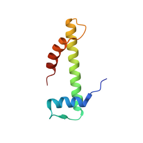
3B0D

Crystal structure of the chicken CENP-T histone fold/CENP-W complex, crystal form II


Experimental Data Snapshot

  • Method: X-RAY DIFFRACTION
  • Resolution: 2.20 Å
  • R-Value Free: 0.261 
  • R-Value Work: 0.203 
  • R-Value Observed: 0.208 

wwPDB Validation   3D Report Full Report


This is version 1.1 of the entry. See complete history


Literature

CENP-T-W-S-X Forms a Unique Centromeric Chromatin Structure with a Histone-like Fold.

Nishino, T.Takeuchi, K.Gascoigne, K.E.Suzuki, A.Hori, T.Oyama, T.Morikawa, K.Cheeseman, I.M.Fukagawa, T.

(2012) Cell 148: 487-501

  • DOI: 10.1016/j.cell.2011.11.061
  • Primary Citation of Related Structures:  
    3B0B, 3B0C, 3B0D, 3VH5, 3VH6

  • PubMed Abstract: 
  • The multiprotein kinetochore complex must assemble at a specific site on each chromosome to achieve accurate chromosome segregation. Defining the nature of the DNA-protein interactions that specify the position of the kinetochore and provide a scaffold for kinetochore formation remain key goals ...

    The multiprotein kinetochore complex must assemble at a specific site on each chromosome to achieve accurate chromosome segregation. Defining the nature of the DNA-protein interactions that specify the position of the kinetochore and provide a scaffold for kinetochore formation remain key goals. Here, we demonstrate that the centromeric histone-fold-containing CENP-T-W and CENP-S-X complexes coassemble to form a stable CENP-T-W-S-X heterotetramer. High-resolution structural analysis of the individual complexes and the heterotetramer reveals similarity to other histone fold-containing complexes including canonical histones within a nucleosome. The CENP-T-W-S-X heterotetramer binds to and supercoils DNA. Mutants designed to compromise heterotetramerization or the DNA-protein contacts around the heterotetramer strongly reduce the DNA binding and supercoiling activities in vitro and compromise kinetochore assembly in vivo. These data suggest that the CENP-T-W-S-X complex forms a unique nucleosome-like structure to generate contacts with DNA, extending the "histone code" beyond canonical nucleosome proteins.


    Organizational Affiliation

    Department of Molecular Genetics, National Institute of Genetics and The Graduate University for Advanced Studies (SOKENDAI), Mishima, Shizuoka 411-8540, Japan.


Macromolecules
Find similar proteins by:  (by identity cutoff)  |  3D Structure
Entity ID: 1
MoleculeChainsSequence LengthOrganismDetailsImage
Centromere protein TC [auth B],
A [auth T]
111Gallus gallusMutation(s): 0 
Gene Names: CENPT
UniProt
Find proteins for F1NPG5 (Gallus gallus)
Explore F1NPG5 
Go to UniProtKB:  F1NPG5
Entity Groups  
Sequence Clusters30% Identity50% Identity70% Identity90% Identity95% Identity100% Identity
UniProt GroupF1NPG5
Protein Feature View
Expand
  • Reference Sequence
Find similar proteins by:  (by identity cutoff)  |  3D Structure
Entity ID: 2
MoleculeChainsSequence LengthOrganismDetailsImage
Centromere protein WD [auth C],
B [auth W]
76Gallus gallusMutation(s): 0 
Gene Names: CENPW
UniProt
Find proteins for P0DJH6 (Gallus gallus)
Explore P0DJH6 
Go to UniProtKB:  P0DJH6
Entity Groups  
Sequence Clusters30% Identity50% Identity70% Identity90% Identity95% Identity100% Identity
UniProt GroupP0DJH6
Protein Feature View
Expand
  • Reference Sequence
Small Molecules
Experimental Data & Validation

Experimental Data

  • Method: X-RAY DIFFRACTION
  • Resolution: 2.20 Å
  • R-Value Free: 0.261 
  • R-Value Work: 0.203 
  • R-Value Observed: 0.208 
  • Space Group: P 31 2 1
Unit Cell:
Length ( Å )Angle ( ˚ )
a = 54.985α = 90
b = 54.985β = 90
c = 235.912γ = 120
Software Package:
Software NamePurpose
HKL-2000data collection
PHASERphasing
PHENIXrefinement
HKL-2000data reduction
HKL-2000data scaling

Structure Validation

View Full Validation Report


Entry History 

Deposition Data

Revision History  (Full details and data files)

  • Version 1.0: 2012-03-07
    Type: Initial release
  • Version 1.1: 2017-10-11
    Changes: Refinement description