3AK5 | pdb_00003ak5

Hemoglobin protease (Hbp) passenger missing domain-2


Experimental Data Snapshot

  • Method: X-RAY DIFFRACTION
  • Resolution: 2.20 Å
  • R-Value Free: 
    0.280 (Depositor), 0.280 (DCC) 
  • R-Value Work: 
    0.226 (Depositor), 0.226 (DCC) 
  • R-Value Observed: 
    0.226 (Depositor) 

Starting Model: experimental
View more details

wwPDB Validation 3D Report Full Report

Validation slider image for 3AK5

This is version 1.3 of the entry. See complete history

Literature

Role of domains within the autotransporter Hbp/Tsh

Nishimura, K.Yoon, Y.-H.Kurihara, A.Unzai, S.Luirink, J.Park, S.-Y.Tame, J.R.H.

(2010) Acta Crystallogr D Biol Crystallogr 66: 1295-1300

  • DOI: https://doi.org/10.1107/S0907444910036966
  • Primary Citation Related Structures: 
    3AK5

  • PubMed Abstract: 

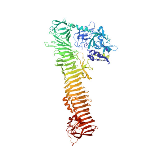
    The autotransporter Tsh (temperature-sensitive haemagglutinin) secreted by avian pathogenic Escherichia coli was reported in 1994 and the almost identical Hbp (haemoglobin protease) was discovered some years later in isolates from patients suffering from peritoneal abscesses. However, the function of the protein remains uncertain. The crystal structure of Hbp shows that the protein carries a serine protease domain (domain 1) and a small domain of 75 residues called domain 2 which is inserted into the long β-helix characteristic of autotransporter passenger proteins. In this paper, domain 1 is shown to bind calcium, although metal ions binding to this site do not seem to regulate protease activity. Tsh has been reported to bind red cells and components of the extracellular matrix, but it is demonstrated that these properties are not a consequence of the presence of domain 2.


  • Organizational Affiliation
    • Yokohama City University, Suehiro 1-7-29, Tsurumi, Yokohama 230-0045, Japan.

Macromolecule Content 

  • Total Structure Weight: 415.06 kDa 
  • Atom Count: 30,307 
  • Modeled Residue Count: 3,840 
  • Deposited Residue Count: 3,900 
  • Unique protein chains: 1

Macromolecules

Find similar proteins by:|  3D Structure
Entity ID: 1
MoleculeChains  Sequence LengthOrganismDetailsImage
Hemoglobin-binding protease hbp
A, B, C, D
975Escherichia coliMutation(s): 0 
Gene Names: HBP
EC: 3.4.21
UniProt
Find proteins for O88093 (Escherichia coli)
Explore O88093 
Go to UniProtKB:  O88093
Entity Groups
Sequence Clusters30% Identity50% Identity70% Identity90% Identity95% Identity100% Identity
UniProt GroupO88093
Sequence Annotations
Expand
Reference Sequence

Experimental Data & Validation

Experimental Data

  • Method: X-RAY DIFFRACTION
  • Resolution: 2.20 Å
  • R-Value Free:  0.280 (Depositor), 0.280 (DCC) 
  • R-Value Work:  0.226 (Depositor), 0.226 (DCC) 
  • R-Value Observed: 0.226 (Depositor) 
Space Group: C 1 2 1
Unit Cell:
Length ( Å )Angle ( ˚ )
a = 292.803α = 90
b = 53.936β = 107.32
c = 335.953γ = 90
Software Package:
Software NamePurpose
ADSCdata collection
EPMRphasing
REFMACrefinement
HKL-2000data reduction
SCALEPACKdata scaling

Structure Validation

View Full Validation Report


Entry History 

Deposition Data

Revision History  (Full details and data files)

  • Version 1.0: 2010-10-06
    Type: Initial release
  • Version 1.1: 2011-07-13
    Changes: Version format compliance
  • Version 1.2: 2017-08-09
    Changes: Advisory, Refinement description, Source and taxonomy
  • Version 1.3: 2023-11-01
    Changes: Advisory, Data collection, Database references, Derived calculations, Refinement description