3AI7

Crystal Structure of Bifidobacterium Longum Phosphoketolase


Experimental Data Snapshot

  • Method: X-RAY DIFFRACTION
  • Resolution: 2.20 Å
  • R-Value Free: 0.206 
  • R-Value Work: 0.147 
  • R-Value Observed: 0.150 

wwPDB Validation   3D Report Full Report


Ligand Structure Quality Assessment 


This is version 1.4 of the entry. See complete history


Literature

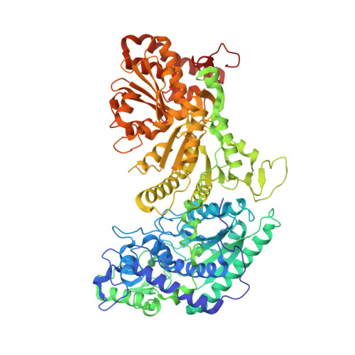
Crystal structure of Bifidobacterium Longum phosphoketolase; key enzyme for glucose metabolism in Bifidobacterium

Takahashi, K.Tagami, U.Shimba, N.Kashiwagi, T.Ishikawa, K.Suzuki, E.

(2010) FEBS Lett 584: 3855-3861

  • DOI: https://doi.org/10.1016/j.febslet.2010.07.043
  • Primary Citation of Related Structures:  
    3AI7

  • PubMed Abstract: 

    The crystal structure of Bifidobacterium longum phosphoketolase, a thiamine diphosphate (TPP) dependent enzyme, has been determined at 2.2A resolution. The enzyme is a dimer with the active sites located at the interface between the two identical subunits with molecular mass of 92.5 kDa. The bound TPP is almost completely shielded from solvent except for the catalytically important C2-carbon of the thiazolium ring, which can be accessed by a substrate sugar through a narrow funnel-shaped channel. In silico docking studies of B. longum phosphoketolase with its substrate enable us to propose a model for substrate binding.


  • Organizational Affiliation

    Institute of Life Sciences, Ajinomoto Co., Inc., 1-1 Suzuki-cho, Kawasaki-ku, Kawasaki, Japan.


Macromolecules
Find similar proteins by:  (by identity cutoff)  |  3D Structure
Entity ID: 1
MoleculeChains Sequence LengthOrganismDetailsImage
Xylulose-5-phosphate/fructose-6-phosphate phosphoketolase
A, B, C, D, E
A, B, C, D, E, F, G, H
831Bifidobacterium longumMutation(s): 0 
EC: 4.1.2.22
UniProt
Find proteins for Q6R2Q7 (Bifidobacterium longum)
Explore Q6R2Q7 
Go to UniProtKB:  Q6R2Q7
Entity Groups  
Sequence Clusters30% Identity50% Identity70% Identity90% Identity95% Identity100% Identity
UniProt GroupQ6R2Q7
Sequence Annotations
Expand
  • Reference Sequence
Small Molecules
Ligands 2 Unique
IDChains Name / Formula / InChI Key2D Diagram3D Interactions
TPP
Query on TPP

Download Ideal Coordinates CCD File 
I [auth A]
K [auth B]
M [auth C]
O [auth D]
Q [auth E]
I [auth A],
K [auth B],
M [auth C],
O [auth D],
Q [auth E],
S [auth F],
U [auth G],
W [auth H]
THIAMINE DIPHOSPHATE
C12 H19 N4 O7 P2 S
AYEKOFBPNLCAJY-UHFFFAOYSA-O
CA
Query on CA

Download Ideal Coordinates CCD File 
J [auth A]
L [auth B]
N [auth C]
P [auth D]
R [auth E]
J [auth A],
L [auth B],
N [auth C],
P [auth D],
R [auth E],
T [auth F],
V [auth G],
X [auth H]
CALCIUM ION
Ca
BHPQYMZQTOCNFJ-UHFFFAOYSA-N
Experimental Data & Validation

Experimental Data

  • Method: X-RAY DIFFRACTION
  • Resolution: 2.20 Å
  • R-Value Free: 0.206 
  • R-Value Work: 0.147 
  • R-Value Observed: 0.150 
  • Space Group: P 1 21 1
Unit Cell:
Length ( Å )Angle ( ˚ )
a = 142.26α = 90
b = 184.41β = 97.31
c = 157.44γ = 90
Software Package:
Software NamePurpose
XSCALEdata scaling
AMoREphasing
REFMACrefinement
PDB_EXTRACTdata extraction

Structure Validation

View Full Validation Report


Ligand Structure Quality Assessment 


Entry History 

Deposition Data

Revision History  (Full details and data files)

  • Version 1.0: 2010-09-15
    Type: Initial release
  • Version 1.1: 2011-07-13
    Changes: Version format compliance
  • Version 1.2: 2014-01-29
    Changes: Database references
  • Version 1.3: 2024-03-13
    Changes: Data collection, Database references, Derived calculations
  • Version 1.4: 2024-04-03
    Changes: Refinement description