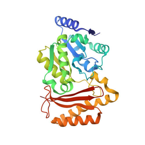
3AG6

Crystal Structure of Pantothenate Synthetase from Staphylococcus aureus in complex with pantoyl adenylate


Experimental Data Snapshot

  • Method: X-RAY DIFFRACTION
  • Resolution: 1.85 Å
  • R-Value Free: 0.228 
  • R-Value Work: 0.201 
  • R-Value Observed: 0.201 

wwPDB Validation   3D Report Full Report


Ligand Structure Quality Assessment 


This is version 1.3 of the entry. See complete history


Literature

Substrate-induced closing of the active site revealed by the crystal structure of pantothenate synthetase from Staphylococcus aureus.

Satoh, A.Konishi, S.Tamura, H.Stickland, H.G.Whitney, H.M.Smith, A.G.Matsumura, H.Inoue, T.

(2010) Biochemistry 49: 6400-6410

  • DOI: https://doi.org/10.1021/bi1004206
  • Primary Citation of Related Structures:  
    3AG5, 3AG6

  • PubMed Abstract: 

    Pantothenate synthetase (PS, EC 6.3.2.1) is the last enzyme in the pantothenate biosynthesis pathway, a metabolic pathway identified as a potential target for new antimicrobials. PS catalyzes the ATP-dependent condensation of pantoate and beta-alanine to form pantothenate. Here we report the overexpression, purification, enzyme assay, and tertiary structure of PS from Staphylococcus aureus. PS activity was experimentally confirmed, indicating a k(cat) value comparable to those of enzymes from other organisms. The structures of the apoenzyme and the reaction intermediate (pantoyl adenylate; PA) complex were determined by X-ray crystallography to resolutions of 2.5 and 1.85 A, respectively. Structural analysis indicated that the apoenzyme adopts an open and relatively mobile structure, while the complex structure is closed and entirely rigid. Structural comparison of the apoenzyme and the complex revealed how S. aureus PS undergoes open/close conformational change, and also determined the key interactions with the adenine ring of PA for a hinge bending domain closure. In the complex structure, PA and acetate are bound in the active site. We suggest that the acetate mimics the substrate beta-alanine. Therefore, the complex structure seems to represent a catalytic state poised for in-line nucleophilic attack on PA. These data also offer an alternative strategy for designing novel compounds that selectively inhibit PS activity.


  • Organizational Affiliation

    Department of Applied Chemistry, Graduate School of Engineering, Osaka University, 2-1 Yamadaoka, Suita, Osaka, Japan.


Macromolecules
Find similar proteins by:  (by identity cutoff)  |  3D Structure
Entity ID: 1
MoleculeChains Sequence LengthOrganismDetailsImage
Pantothenate synthetase
A, B
283Staphylococcus aureus subsp. aureus NCTC 8325Mutation(s): 0 
Gene Names: panCSAOUHSC_02918
EC: 6.3.2.1
UniProt
Find proteins for Q2FV22 (Staphylococcus aureus (strain NCTC 8325 / PS 47))
Explore Q2FV22 
Go to UniProtKB:  Q2FV22
Entity Groups  
Sequence Clusters30% Identity50% Identity70% Identity90% Identity95% Identity100% Identity
UniProt GroupQ2FV22
Sequence Annotations
Expand
  • Reference Sequence
Small Molecules
Ligands 3 Unique
IDChains Name / Formula / InChI Key2D Diagram3D Interactions
PAJ
Query on PAJ

Download Ideal Coordinates CCD File 
C [auth A],
I [auth B]
PANTOYL ADENYLATE
C16 H24 N5 O10 P
GDPVENOGSVMRJL-FSRKKXLISA-N
PG4
Query on PG4

Download Ideal Coordinates CCD File 
E [auth A]
F [auth A]
G [auth A]
H [auth A]
K [auth B]
E [auth A],
F [auth A],
G [auth A],
H [auth A],
K [auth B],
L [auth B],
M [auth B],
N [auth B]
TETRAETHYLENE GLYCOL
C8 H18 O5
UWHCKJMYHZGTIT-UHFFFAOYSA-N
ACY
Query on ACY

Download Ideal Coordinates CCD File 
D [auth A],
J [auth B]
ACETIC ACID
C2 H4 O2
QTBSBXVTEAMEQO-UHFFFAOYSA-N
Experimental Data & Validation

Experimental Data

  • Method: X-RAY DIFFRACTION
  • Resolution: 1.85 Å
  • R-Value Free: 0.228 
  • R-Value Work: 0.201 
  • R-Value Observed: 0.201 
  • Space Group: P 1 21 1
Unit Cell:
Length ( Å )Angle ( ˚ )
a = 46.026α = 90
b = 91.453β = 103.11
c = 87.342γ = 90
Software Package:
Software NamePurpose
HKL-2000data collection
MOLREPphasing
CNSrefinement
HKL-2000data reduction
HKL-2000data scaling

Structure Validation

View Full Validation Report


Ligand Structure Quality Assessment 


Entry History 

Deposition Data

Revision History  (Full details and data files)

  • Version 1.0: 2010-07-21
    Type: Initial release
  • Version 1.1: 2011-07-13
    Changes: Version format compliance
  • Version 1.2: 2013-09-04
    Changes: Database references
  • Version 1.3: 2024-03-13
    Changes: Data collection, Database references, Derived calculations