3A9J | pdb_00003a9j

Crystal structure of the mouse TAB2-NZF in complex with Lys63-linked di-ubiquitin


Experimental Data Snapshot

  • Method: X-RAY DIFFRACTION
  • Resolution: 1.18 Å
  • R-Value Free: 
    0.191 (Depositor), 0.189 (DCC) 
  • R-Value Work: 
    0.164 (Depositor), 0.163 (DCC) 
  • R-Value Observed: 
    0.166 (Depositor) 

wwPDB Validation 3D Report Full Report

Validation slider image for 3A9J

This is version 1.3 of the entry. See complete history

Literature

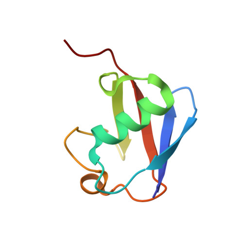
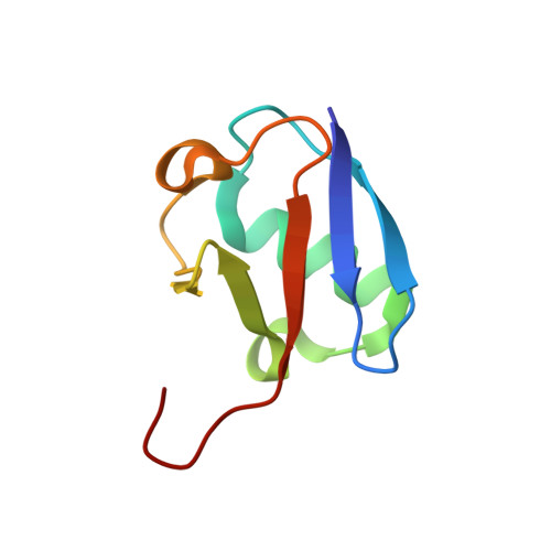
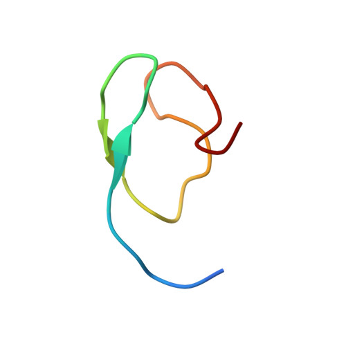
Structural basis for specific recognition of Lys 63-linked polyubiquitin chains by NZF domains of TAB2 and TAB3

Sato, Y.Yoshikawa, A.Yamashita, M.Yamagata, A.Fukai, S.

(2009) EMBO J 28: 3903-3909

  • DOI: https://doi.org/10.1038/emboj.2009.345
  • Primary Citation Related Structures: 
    3A9J, 3A9K

  • PubMed Abstract: 

    TAB2 and TAB3 activate the Jun N-terminal kinase and nuclear factor-kappaB pathways through the specific recognition of Lys 63-linked polyubiquitin chains by its Npl4 zinc-finger (NZF) domain. Here we report crystal structures of the TAB2 and TAB3 NZF domains in complex with Lys 63-linked diubiquitin at 1.18 and 1.40 A resolutions, respectively. Both NZF domains bind to the distal ubiquitin through a conserved Thr-Phe dipeptide that has been shown to be important for the interaction of the NZF domain of Npl4 with monoubiquitin. In contrast, a surface specific to TAB2 and TAB3 binds the proximal ubiquitin. Both the distal and proximal binding sites of the TAB2 and TAB3 NZF domains recognize the Ile 44-centred hydrophobic patch on ubiquitin but do not interact with the Lys 63-linked isopeptide bond. Mutagenesis experiments show that both binding sites are required to enable binding of Lys 63-linked diubiquitin. We therefore propose a mechanism for the recognition of Lys 63-linked polyubiquitin chains by TAB2 and TAB3 NZF domains in which diubiquitin units are specifically recognized by a single NZF domain.


  • Organizational Affiliation
    • Structural Biology Laboratory, Life Science Division, Synchrotron Radiation Research Organization, Institute of Molecular and Cellular Biosciences, The University of Tokyo, Tokyo, Japan.

Macromolecule Content 

  • Total Structure Weight: 21.22 kDa 
  • Atom Count: 1,706 
  • Modeled Residue Count: 185 
  • Deposited Residue Count: 187 
  • Unique protein chains: 3

Macromolecules

Find similar proteins by:|  3D Structure
Entity ID: 1
MoleculeChains  Sequence LengthOrganismDetailsImage
Ubiquitin76Mus musculusMutation(s): 1 
Gene Names: Ubiquitin
UniProt & NIH Common Fund Data Resources
Find proteins for P0CG50 (Mus musculus)
Explore P0CG50 
Go to UniProtKB:  P0CG50
IMPC:  MGI:98889
Entity Groups
Sequence Clusters30% Identity50% Identity70% Identity90% Identity95% Identity100% Identity
UniProt GroupP0CG50
Sequence Annotations
Expand
Reference Sequence
Find similar proteins by:|  3D Structure
Entity ID: 2
MoleculeChains  Sequence LengthOrganismDetailsImage
Ubiquitin77Mus musculusMutation(s): 1 
Gene Names: Ubiquitin
UniProt & NIH Common Fund Data Resources
Find proteins for P0CG50 (Mus musculus)
Explore P0CG50 
Go to UniProtKB:  P0CG50
IMPC:  MGI:98889
Entity Groups
Sequence Clusters30% Identity50% Identity70% Identity90% Identity95% Identity100% Identity
UniProt GroupP0CG50
Sequence Annotations
Expand
Reference Sequence
Find similar proteins by:|  3D Structure
Entity ID: 3
MoleculeChains  Sequence LengthOrganismDetailsImage
Mitogen-activated protein kinase kinase kinase 7-interacting protein 234Mus musculusMutation(s): 0 
Gene Names: TAB2 (AMINO ACIDS 665 - 693)
UniProt
Find proteins for Q99K90 (Mus musculus)
Explore Q99K90 
Go to UniProtKB:  Q99K90
Entity Groups
Sequence Clusters30% Identity50% Identity70% Identity90% Identity95% Identity100% Identity
UniProt GroupQ99K90
Sequence Annotations
Expand
Reference Sequence

Small Molecules

Ligands 1 Unique
IDChains Name / Formula / InChI Key2D Diagram3D Interactions
ZN

Query on ZN


Download:Ideal Coordinates CCD File
D [auth C]ZINC ION
Zn
PTFCDOFLOPIGGS-UHFFFAOYSA-N

Experimental Data & Validation

Experimental Data

  • Method: X-RAY DIFFRACTION
  • Resolution: 1.18 Å
  • R-Value Free:  0.191 (Depositor), 0.189 (DCC) 
  • R-Value Work:  0.164 (Depositor), 0.163 (DCC) 
  • R-Value Observed: 0.166 (Depositor) 
Space Group: P 21 21 21
Unit Cell:
Length ( Å )Angle ( ˚ )
a = 30.085α = 90
b = 71.478β = 90
c = 72.029γ = 90
Software Package:
Software NamePurpose
ADSCdata collection
SHARPphasing
REFMACrefinement
HKL-2000data reduction
HKL-2000data scaling

Structure Validation

View Full Validation Report


Entry History 

Deposition Data

Revision History  (Full details and data files)

  • Version 1.0: 2009-12-08
    Type: Initial release
  • Version 1.1: 2011-07-13
    Changes: Version format compliance
  • Version 1.2: 2021-11-10
    Changes: Database references, Derived calculations
  • Version 1.3: 2024-10-30
    Changes: Data collection, Structure summary