3ZXB | pdb_00003zxb

Wild type single insulin-like growth factor binding domain protein (SIBD-1) from Cupiennius salei


Experimental Data Snapshot

  • Method: X-RAY DIFFRACTION
  • Resolution: 2.55 Å
  • R-Value Free: 
    0.250 (Depositor), 0.240 (DCC) 
  • R-Value Work: 
    0.224 (Depositor), 0.230 (DCC) 
  • R-Value Observed: 
    0.226 (Depositor) 

Starting Model: experimental
View more details

wwPDB Validation   3D Report Full Report


This is version 1.3 of the entry. See complete history


Literature

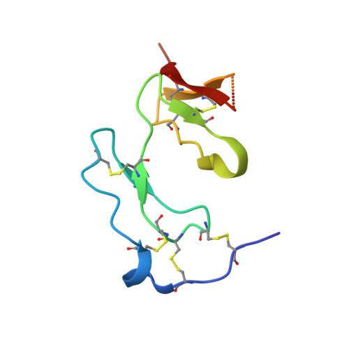
Structural and Biochemical Characterization of Native and Recombinant Single Insulin-Like Growth Factor-Binding Domain Protein (Sibd-1) from the Central American Hunting Spider Cupiennius Salei (Ctenidae).

Trachsel, C.Widmer, C.Kampfer, U.Buhr, C.Baumann, T.Kuhn-Nentwig, L.Schurch, S.Schaller, J.Baumann, U.

(2012) Proteins 80: 2323

  • DOI: https://doi.org/10.1002/prot.24119
  • Primary Citation of Related Structures:  
    3ZXB, 3ZXC

  • PubMed Abstract: 

    Cupiennius salei single insulin-like growth factor binding domain protein (SIBD-1) is an 8.6 kDa Cys-, Pro-, and Gly-rich protein, discovered in the hemocytes of the Central American hunting spider Cupiennius salei. SIBD-1 exhibits high sequence similarity to the N-terminal domain of the insulin-like growth factor-binding protein superfamily and has been reported to play an important role in the spider's immune system. Here, the recombinant expression and the elucidation of the three-dimensional structure of recombinant SIBD-1 and the characterization of the sugar moiety at Thr2 of native SIBD-1 is described in detail.


  • Organizational Affiliation
    • Department of Chemistry and Biochemistry, University of Bern, Bern, Switzerland.

Macromolecules
Find similar proteins by:  (by identity cutoff)  |  3D Structure
Entity ID: 1
MoleculeChains Sequence LengthOrganismDetailsImage
SINGLE INSULIN-LIKE GROWTH FACTOR-BINDING DOMAIN PROTEIN-1
A, B, C, D
78Cupiennius saleiMutation(s): 0 
UniProt
Find proteins for G4V4F9 (Cupiennius salei)
Explore G4V4F9 
Go to UniProtKB:  G4V4F9
Entity Groups  
Sequence Clusters30% Identity50% Identity70% Identity90% Identity95% Identity100% Identity
UniProt GroupG4V4F9
Sequence Annotations
Expand
  • Reference Sequence
Experimental Data & Validation

Experimental Data

  • Method: X-RAY DIFFRACTION
  • Resolution: 2.55 Å
  • R-Value Free:  0.250 (Depositor), 0.240 (DCC) 
  • R-Value Work:  0.224 (Depositor), 0.230 (DCC) 
  • R-Value Observed: 0.226 (Depositor) 
Space Group: P 32 2 1
Unit Cell:
Length ( Å )Angle ( ˚ )
a = 62.247α = 90
b = 62.247β = 90
c = 169.297γ = 120
Software Package:
Software NamePurpose
PHENIXrefinement
XDSdata reduction
XSCALEdata scaling
PHASERphasing

Structure Validation

View Full Validation Report


Entry History 

Deposition Data

Revision History  (Full details and data files)

  • Version 1.0: 2012-06-06
    Type: Initial release
  • Version 1.1: 2012-08-08
    Changes: Database references
  • Version 1.2: 2023-12-20
    Changes: Data collection, Database references, Other, Refinement description
  • Version 1.3: 2024-11-20
    Changes: Structure summary