3ZWN

Crystal structure of Aplysia cyclase complexed with substrate NGD and product cGDPR


Experimental Data Snapshot

  • Method: X-RAY DIFFRACTION
  • Resolution: 1.80 Å
  • R-Value Free: 0.233 
  • R-Value Work: 0.176 
  • R-Value Observed: 0.179 

wwPDB Validation   3D Report Full Report


Ligand Structure Quality Assessment 


This is version 1.2 of the entry. See complete history


Literature

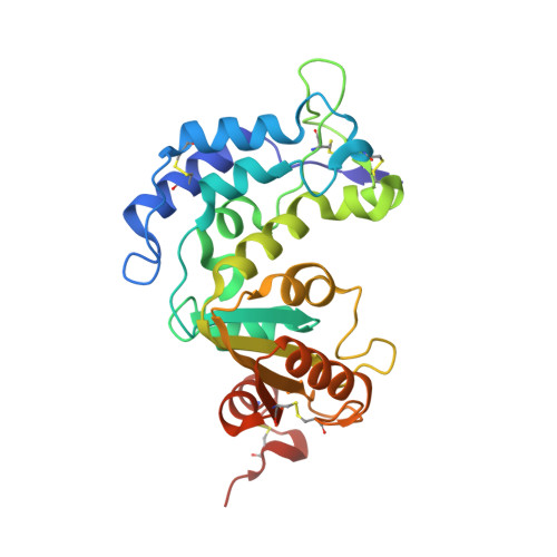
Structural Studies of Intermediates Along the Cyclization Pathway of Aplysia Adp-Ribosyl Cyclase.

Kotaka, M.Graeff, R.Chen, Z.Zhang, L.H.Lee, H.C.Hao, Q.

(2012) J Mol Biol 415: 514

  • DOI: https://doi.org/10.1016/j.jmb.2011.11.022
  • Primary Citation of Related Structures:  
    3ZWM, 3ZWN, 3ZWO, 3ZWP, 3ZWV, 3ZWW

  • PubMed Abstract: 

    Cyclic ADP-ribose (cADPR) is a calcium messenger that can mobilize intracellular Ca²⁺ stores and activate Ca²⁺ influx to regulate a wide range of physiological processes. Aplysia cyclase is the first member of the ADP-ribosyl cyclases identified to catalyze the cyclization of NAD⁺ into cADPR. The catalysis involves a two-step reaction, the elimination of the nicotinamide ring and the cyclization of the intermediate resulting in the covalent attachment of the purine ring to the terminal ribose. Aplysia cyclase exhibits a high degree of leniency towards the purine base of its substrate, and the cyclization reaction takes place at either the N1- or the N7-position of the purine ring. To decipher the mechanism of cyclization in Aplysia cyclase, we used a crystallization setup with multiple Aplysia cyclase molecules present in the asymmetric unit. With the use of natural substrates and analogs, not only were we able to capture multiple snapshots during enzyme catalysis resulting in either N1 or N7 linkage of the purine ring to the terminal ribose, we were also able to observe, for the first time, the cyclized products of both N1 and N7 cyclization bound in the active site of Aplysia cyclase.


  • Organizational Affiliation

    Department of Physiology, University of Hong Kong, Hong Kong SAR, China.


Macromolecules
Find similar proteins by:  (by identity cutoff)  |  3D Structure
Entity ID: 1
MoleculeChains Sequence LengthOrganismDetailsImage
ADP-RIBOSYL CYCLASE
A, B
260Aplysia californicaMutation(s): 0 
EC: 3.2.2.5
UniProt
Find proteins for P29241 (Aplysia californica)
Explore P29241 
Go to UniProtKB:  P29241
Entity Groups  
Sequence Clusters30% Identity50% Identity70% Identity90% Identity95% Identity100% Identity
UniProt GroupP29241
Sequence Annotations
Expand
  • Reference Sequence
Small Molecules
Ligands 2 Unique
IDChains Name / Formula / InChI Key2D Diagram3D Interactions
NGD
Query on NGD

Download Ideal Coordinates CCD File 
D [auth B]3-(AMINOCARBONYL)-1-[(2R,3R,4S,5R)-5-({[(S)-{[(S)-{[(2R,3S,4R,5R)-5-(2-AMINO-6-OXO-1,6-DIHYDRO-9H-PURIN-9-YL)-3,4-DIHYD ROXYTETRAHYDROFURAN-2-YL]METHOXY}(HYDROXY)PHOSPHORYL]OXY}(HYDROXY)PHOSPHORYL]OXY}METHYL)-3,4-DIHYDROXYTETRAHYDROFURAN-2- YL]PYRIDINIUM
C21 H28 N7 O15 P2
NCFRZZVNRVEQJK-NAJQWHGHSA-O
CGR
Query on CGR

Download Ideal Coordinates CCD File 
C [auth A]CYCLIC GUANOSINE DIPHOSPHATE-RIBOSE
C15 H23 N5 O14 P2
XBIQPWWXYHHVOT-AIKIOGRUSA-N
Experimental Data & Validation

Experimental Data

  • Method: X-RAY DIFFRACTION
  • Resolution: 1.80 Å
  • R-Value Free: 0.233 
  • R-Value Work: 0.176 
  • R-Value Observed: 0.179 
  • Space Group: P 1 21 1
Unit Cell:
Length ( Å )Angle ( ˚ )
a = 69.494α = 90
b = 57.852β = 100.94
c = 71.303γ = 90
Software Package:
Software NamePurpose
REFMACrefinement
HKL-2000data reduction
HKL-2000data scaling
MOLREPphasing

Structure Validation

View Full Validation Report


Ligand Structure Quality Assessment 


Entry History 

Deposition Data

Revision History  (Full details and data files)

  • Version 1.0: 2011-11-30
    Type: Initial release
  • Version 1.1: 2012-01-25
    Changes: Other
  • Version 1.2: 2023-12-20
    Changes: Data collection, Database references, Derived calculations, Other, Refinement description, Structure summary