3ZW0

Structure of BambL lectin from Burkholderia ambifaria


Experimental Data Snapshot

  • Method: X-RAY DIFFRACTION
  • Resolution: 1.60 Å
  • R-Value Free: 0.188 
  • R-Value Work: 0.143 
  • R-Value Observed: 0.146 

wwPDB Validation   3D Report Full Report


Ligand Structure Quality Assessment 


This is version 1.3 of the entry. See complete history


Literature

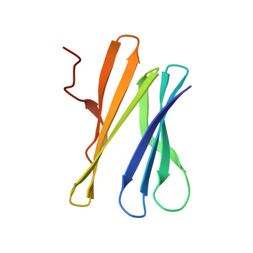
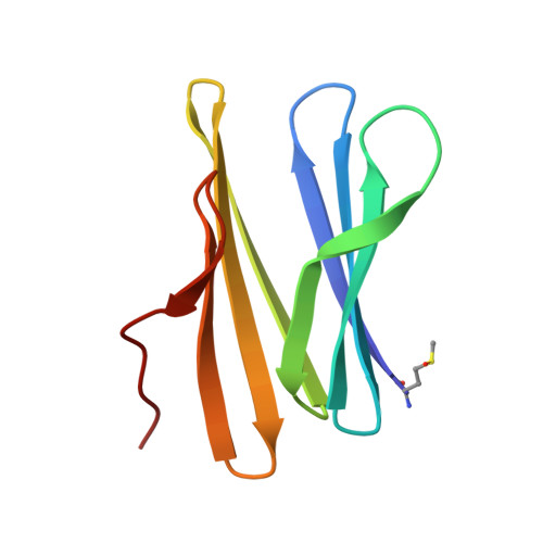
Fucose-Binding Lectin from Opportunistic Pathogen Burkholderia Ambifaria Binds to Both Plant and Human Oligosaccharidic Epitopes.

Audfray, A.Claudinon, J.Abounit, S.Ruvoen-Clouet, N.Larson, G.Smith, D.F.Wimmerova, M.Le Pendu, J.Romer, W.Varrot, A.Imberty, A.

(2012) J Biol Chem 287: 4335

  • DOI: https://doi.org/10.1074/jbc.M111.314831
  • Primary Citation of Related Structures:  
    3ZW0, 3ZW2, 3ZWE, 3ZZV

  • PubMed Abstract: 

    Burkholderia ambifaria is generally associated with the rhizosphere of plants where it has biocontrol effects on other microorganisms. It is also a member of the Burkholderia cepacia complex, a group of closely related bacteria that cause lung infections in immunocompromised patients as well as in patients with granulomatous disease or cystic fibrosis. Our previous work indicated that fucose on human epithelia is a frequent target for lectins and adhesins of lung pathogens (Sulák, O., Cioci, G., Lameignère, E., Balloy, V., Round, A., Gutsche, I., Malinovská, L., Chignard, M., Kosma, P., Aubert, D. F., Marolda, C. L., Valvano, M. A., Wimmerová, M., and Imberty, A. (2011) PLoS Pathog. 7, e1002238). Analysis of the B. ambifaria genome identified BambL as a putative fucose-binding lectin. The 87-amino acid protein was produced recombinantly and demonstrated to bind to fucosylated oligosaccharides with a preference for αFuc1-2Gal epitopes. Crystal structures revealed that it associates as a trimer with two fucose-binding sites per monomer. The overall fold is a six-bladed β-propeller formed by oligomerization as in the Ralstonia solanacearum lectin and not by sequential domains like the fungal fucose lectin from Aleuria aurantia. The affinity of BambL for small fucosylated glycans is very high as demonstrated by microcalorimetry (K(D) < 1 μM). Plant cell wall oligosaccharides and human histo-blood group oligosaccharides H-type 2 and Lewis Y are bound with equivalent efficiency. Binding to artificial glycosphingolipid-containing vesicles, human saliva, and lung tissues confirmed that BambL could recognize a wide spectrum of fucosylated epitopes, albeit with a lower affinity for biological material from nonsecretor individuals.


  • Organizational Affiliation

    Centre de Recherche sur les Macromolécules Végétales (CERMAV)-CNRS, Université Joseph Fourier and Institut de Chimie Moléculaire de Grenoble, 38041 Grenoble, France.


Macromolecules
Find similar proteins by:  (by identity cutoff)  |  3D Structure
Entity ID: 1
MoleculeChains Sequence LengthOrganismDetailsImage
BAMBL LECTIN
A, C
87Burkholderia ambifaria AMMDMutation(s): 0 
UniProt
Find proteins for Q0B4G1 (Burkholderia ambifaria (strain ATCC BAA-244 / AMMD))
Explore Q0B4G1 
Go to UniProtKB:  Q0B4G1
Entity Groups  
Sequence Clusters30% Identity50% Identity70% Identity90% Identity95% Identity100% Identity
UniProt GroupQ0B4G1
Sequence Annotations
Expand
  • Reference Sequence
Find similar proteins by:  (by identity cutoff)  |  3D Structure
Entity ID: 2
MoleculeChains Sequence LengthOrganismDetailsImage
BAMBL LECTIN87Burkholderia ambifaria AMMDMutation(s): 0 
UniProt
Find proteins for Q0B4G1 (Burkholderia ambifaria (strain ATCC BAA-244 / AMMD))
Explore Q0B4G1 
Go to UniProtKB:  Q0B4G1
Entity Groups  
Sequence Clusters30% Identity50% Identity70% Identity90% Identity95% Identity100% Identity
UniProt GroupQ0B4G1
Sequence Annotations
Expand
  • Reference Sequence
Small Molecules
Ligands 1 Unique
IDChains Name / Formula / InChI Key2D Diagram3D Interactions
FUC
Query on FUC

Download Ideal Coordinates CCD File 
D [auth C]alpha-L-fucopyranose
C6 H12 O5
SHZGCJCMOBCMKK-SXUWKVJYSA-N
Modified Residues  1 Unique
IDChains TypeFormula2D DiagramParent
MHO
Query on MHO
B
L-PEPTIDE LINKINGC5 H11 N O3 SMET
Experimental Data & Validation

Experimental Data

  • Method: X-RAY DIFFRACTION
  • Resolution: 1.60 Å
  • R-Value Free: 0.188 
  • R-Value Work: 0.143 
  • R-Value Observed: 0.146 
  • Space Group: P 21 21 21
Unit Cell:
Length ( Å )Angle ( ˚ )
a = 45.11α = 90
b = 47.25β = 90
c = 98.33γ = 90
Software Package:
Software NamePurpose
REFMACrefinement
MOSFLMdata reduction
SCALAdata scaling
PHASERphasing

Structure Validation

View Full Validation Report


Ligand Structure Quality Assessment 


Entry History 

Deposition Data

Revision History  (Full details and data files)

  • Version 1.0: 2011-12-21
    Type: Initial release
  • Version 1.1: 2012-02-15
    Changes: Other
  • Version 1.2: 2020-07-29
    Type: Remediation
    Reason: Carbohydrate remediation
    Changes: Data collection, Derived calculations, Other, Structure summary
  • Version 1.3: 2023-12-20
    Changes: Data collection, Database references, Refinement description, Structure summary