3ZRZ | pdb_00003zrz

Crystal structure of the second and third fibronectin F1 modules in complex with a fragment of Streptococcus pyogenes SfbI-5


Experimental Data Snapshot

  • Method: X-RAY DIFFRACTION
  • Resolution: 1.70 Å
  • R-Value Free: 
    0.222 (Depositor), 0.236 (DCC) 
  • R-Value Work: 
    0.174 (Depositor) 
  • R-Value Observed: 
    0.176 (Depositor) 

Starting Model: experimental
View more details

wwPDB Validation 3D Report Full Report

Validation slider image for 3ZRZ

This is version 1.6 of the entry. See complete history

Literature

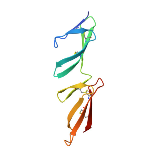
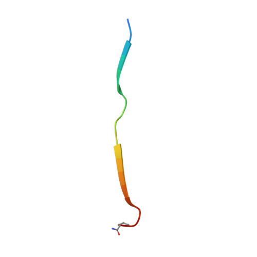
Structural and Functional Analysis of the Tandem Beta-Zipper Interaction of a Streptococcal Protein with Human Fibronectin.

Norris, N.C.Bingham, R.J.Harris, G.Speakman, A.Jones, R.P.O.Leech, A.Turkenburg, J.P.Potts, J.R.

(2011) J Biological Chem 286: 38311

  • DOI: https://doi.org/10.1074/jbc.M111.276592
  • Primary Citation Related Structures: 
    3ZRZ

  • PubMed Abstract: 

    Bacterial fibronectin-binding proteins (FnBPs) contain a large intrinsically disordered region (IDR) that mediates adhesion of bacteria to host tissues, and invasion of host cells, through binding to fibronectin (Fn). These FnBP IDRs consist of Fn-binding repeats (FnBRs) that form a highly extended tandem β-zipper interaction on binding to the N-terminal domain of Fn. Several FnBR residues are highly conserved across bacterial species, and here we investigate their contribution to the interaction. Mutation of these residues to alanine in SfbI-5 (a disordered FnBR from the human pathogen Streptococcus pyogenes) reduced binding, but for each residue the change in free energy of binding was <2 kcal/mol. The structure of an SfbI-5 peptide in complex with the second and third F1 modules from Fn confirms that the conserved FnBR residues play equivalent functional roles across bacterial species. Thus, in SfbI-5, the binding energy for the tandem β-zipper interaction with Fn is distributed across the interface rather than concentrated in a small number of "hot spot" residues that are frequently observed in the interactions of folded proteins. We propose that this might be a common feature of the interactions of IDRs and is likely to pose a challenge for the development of small molecule inhibitors of FnBP-mediated adhesion to and invasion of host cells.


  • Organizational Affiliation
    • Department of Biology, University of York, York, YO10 5DD, United Kingdom.

Macromolecule Content 

  • Total Structure Weight: 24.43 kDa 
  • Atom Count: 1,893 
  • Modeled Residue Count: 218 
  • Deposited Residue Count: 220 
  • Unique protein chains: 2

Macromolecules

Find similar proteins by:|  3D Structure
Entity ID: 1
MoleculeChains  Sequence LengthOrganismDetailsImage
FIBRONECTIN
A, B
90Homo sapiensMutation(s): 0 
UniProt & NIH Common Fund Data Resources
Find proteins for P02751 (Homo sapiens)
Explore P02751 
Go to UniProtKB:  P02751
PHAROS:  P02751
GTEx:  ENSG00000115414 
Entity Groups
Sequence Clusters30% Identity50% Identity70% Identity90% Identity95% Identity100% Identity
UniProt GroupP02751
Sequence Annotations
Expand
Reference Sequence
Find similar proteins by:  Sequence   |   3D Structure  
Entity ID: 2
MoleculeChains  Sequence LengthOrganismDetailsImage
FIBRONECTIN-BINDING PROTEIN
C, D
20Streptococcus pyogenesMutation(s): 2 
UniProt
Find proteins for Q711B0 (Streptococcus pyogenes)
Explore Q711B0 
Go to UniProtKB:  Q711B0
Entity Groups
Sequence Clusters30% Identity50% Identity70% Identity90% Identity95% Identity100% Identity
UniProt GroupQ711B0
Sequence Annotations
Expand
Reference Sequence

Experimental Data & Validation

Experimental Data

  • Method: X-RAY DIFFRACTION
  • Resolution: 1.70 Å
  • R-Value Free:  0.222 (Depositor), 0.236 (DCC) 
  • R-Value Work:  0.174 (Depositor) 
  • R-Value Observed: 0.176 (Depositor) 
Space Group: P 41
Unit Cell:
Length ( Å )Angle ( ˚ )
a = 50.055α = 90
b = 50.055β = 90
c = 93.504γ = 90
Software Package:
Software NamePurpose
REFMACrefinement
MOSFLMdata reduction
SCALAdata scaling
PHASERphasing

Structure Validation

View Full Validation Report


Entry History 

Deposition Data

Revision History  (Full details and data files)

  • Version 1.0: 2011-09-28
    Type: Initial release
  • Version 1.1: 2011-10-19
    Changes: Database references
  • Version 1.2: 2011-11-09
    Changes: Database references, Non-polymer description
  • Version 1.3: 2019-05-08
    Changes: Data collection, Derived calculations, Experimental preparation, Other
  • Version 1.4: 2019-10-23
    Changes: Data collection, Database references, Other
  • Version 1.5: 2023-12-20
    Changes: Data collection, Database references, Derived calculations, Refinement description
  • Version 1.6: 2024-11-06
    Changes: Structure summary