3ZQU

STRUCTURE OF A PROBABLE AROMATIC ACID DECARBOXYLASE


Experimental Data Snapshot

  • Method: X-RAY DIFFRACTION
  • Resolution: 1.50 Å
  • R-Value Free: 0.195 
  • R-Value Work: 0.171 
  • R-Value Observed: 0.173 

wwPDB Validation   3D Report Full Report


Ligand Structure Quality Assessment 


This is version 1.2 of the entry. See complete history


Literature

Structure of Pa4019, a Putative Aromatic Acid Decarboxylase from Pseudomonas Aeruginosa

Kopec, J.Schnell, R.Schneider, G.

(2011) Acta Crystallogr Sect F Struct Biol Cryst Commun 67: 1184

  • DOI: https://doi.org/10.1107/S174430911102923X
  • Primary Citation of Related Structures:  
    3ZQU

  • PubMed Abstract: 

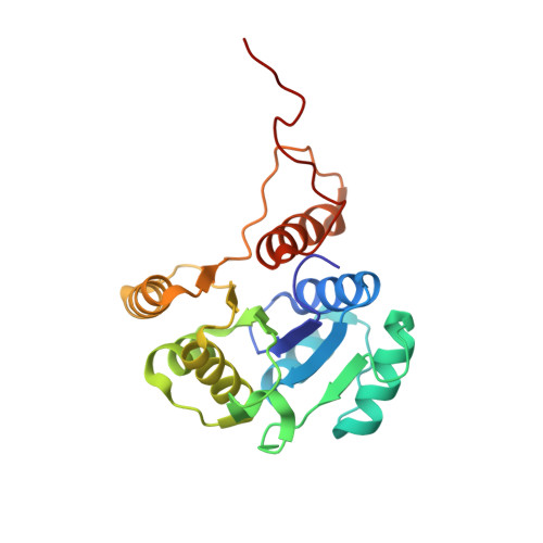
    The ubiX gene (PA4019) of Pseudomonas aeruginosa has been annotated as encoding a putative 3-octaprenyl-4-hydroxybenzoate decarboxylase from the ubiquinone-biosynthesis pathway. Based on a transposon mutagenesis screen, this gene was also implicated as being essential for the survival of this organism. The crystal structure of recombinant UbiX determined to 1.5 Å resolution showed that the protein belongs to the superfamily of homo-oligomeric flavine-containing cysteine decarboxylases. The enzyme assembles into a dodecamer with 23 point symmetry. The subunit displays a typical Rossmann fold and contains one FMN molecule bound at the interface between two subunits.


  • Organizational Affiliation

    Department of Medical Biochemistry and Biophysics, Karolinska Institutet, S-171 77 Stockholm, Sweden.


Macromolecules
Find similar proteins by:  (by identity cutoff)  |  3D Structure
Entity ID: 1
MoleculeChains Sequence LengthOrganismDetailsImage
PROBABLE AROMATIC ACID DECARBOXYLASE209Pseudomonas aeruginosa PAO1Mutation(s): 0 
UniProt
Find proteins for Q9HX08 (Pseudomonas aeruginosa (strain ATCC 15692 / DSM 22644 / CIP 104116 / JCM 14847 / LMG 12228 / 1C / PRS 101 / PAO1))
Explore Q9HX08 
Go to UniProtKB:  Q9HX08
Entity Groups  
Sequence Clusters30% Identity50% Identity70% Identity90% Identity95% Identity100% Identity
UniProt GroupQ9HX08
Sequence Annotations
Expand
  • Reference Sequence
Small Molecules
Ligands 2 Unique
IDChains Name / Formula / InChI Key2D Diagram3D Interactions
FNR
Query on FNR

Download Ideal Coordinates CCD File 
B [auth A]1-DEOXY-1-(7,8-DIMETHYL-2,4-DIOXO-3,4-DIHYDRO-2H-BENZO[G]PTERIDIN-1-ID-10(5H)-YL)-5-O-PHOSPHONATO-D-RIBITOL
C17 H23 N4 O9 P
YTNIXZGTHTVJBW-SCRDCRAPSA-N
SO4
Query on SO4

Download Ideal Coordinates CCD File 
C [auth A],
D [auth A]
SULFATE ION
O4 S
QAOWNCQODCNURD-UHFFFAOYSA-L
Experimental Data & Validation

Experimental Data

  • Method: X-RAY DIFFRACTION
  • Resolution: 1.50 Å
  • R-Value Free: 0.195 
  • R-Value Work: 0.171 
  • R-Value Observed: 0.173 
  • Space Group: F 2 3
Unit Cell:
Length ( Å )Angle ( ˚ )
a = 141.77α = 90
b = 141.77β = 90
c = 141.77γ = 90
Software Package:
Software NamePurpose
REFMACrefinement
MOSFLMdata reduction
SCALAdata scaling
PHASERphasing

Structure Validation

View Full Validation Report


Ligand Structure Quality Assessment 


Entry History 

Deposition Data

Revision History  (Full details and data files)

  • Version 1.0: 2011-11-02
    Type: Initial release
  • Version 1.1: 2012-03-28
    Changes: Other
  • Version 1.2: 2023-12-20
    Changes: Data collection, Database references, Derived calculations, Other, Refinement description