Structural Basis for DNA Recognition and Loading Into a Viral Packaging Motor.
Buttner, C.R., Chechik, M., Ortiz-Lombardia, M., Smits, C., Ebong, I.O., Chechik, V., Jeschke, G., Dykeman, E., Benini, S., Robinson, C.V., Alonso, J.C., Antson, A.A.(2012) Proc Natl Acad Sci U S A 109: 811
- PubMed: 22207627 Search on PubMedSearch on PubMed Central
- DOI: https://doi.org/10.1073/pnas.1110270109
- Primary Citation Related Structures:
3ZQM, 3ZQN, 3ZQO, 3ZQP, 3ZQQ - PubMed Abstract:
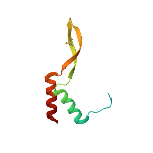
Genome packaging into preformed viral procapsids is driven by powerful molecular motors. The small terminase protein is essential for the initial recognition of viral DNA and regulates the motor's ATPase and nuclease activities during DNA translocation. The crystal structure of a full-length small terminase protein from the Siphoviridae bacteriophage SF6, comprising the N-terminal DNA binding, the oligomerization core, and the C-terminal β-barrel domains, reveals a nine-subunit circular assembly in which the DNA-binding domains are arranged around the oligomerization core in a highly flexible manner. Mass spectrometry analysis and four further crystal structures show that, although the full-length protein exclusively forms nine-subunit assemblies, protein constructs missing the C-terminal β-barrel form both nine-subunit and ten-subunit assemblies, indicating the importance of the C terminus for defining the oligomeric state. The mechanism by which a ring-shaped small terminase oligomer binds viral DNA has not previously been elucidated. Here, we probed binding in vitro by using EPR and surface plasmon resonance experiments, which indicated that interaction with DNA is mediated exclusively by the DNA-binding domains and suggested a nucleosome-like model in which DNA binds around the outside of the protein oligomer.
- York Structural Biology Laboratory, Department of Chemistry, University of York, York, YO10 5DD, United Kingdom.
Organizational Affiliation: