Structural and Genetic Analyses Reveal the Protein Sepf as a New Membrane Anchor for the Z Ring.
Duman, R., Ishikawa, S., Celik, I., Strahl, H., Ogasawara, N., Troc, P., Lowe, J., Hamoen, L.W.(2013) Proc Natl Acad Sci U S A 110: E4601
- PubMed: 24218584 Search on PubMedSearch on PubMed Central
- DOI: https://doi.org/10.1073/pnas.1313978110
- Primary Citation Related Structures:
3ZIE, 3ZIG, 3ZIH, 3ZII - PubMed Abstract:
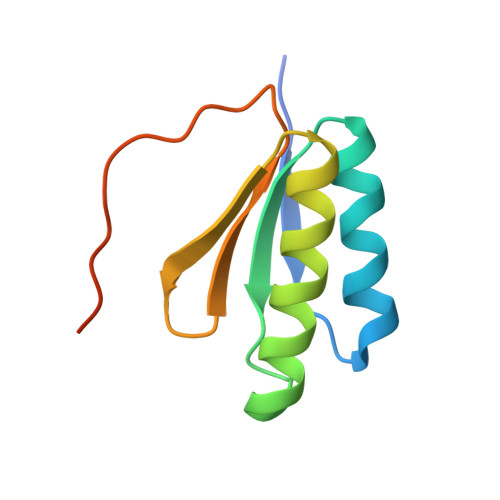
A key step in bacterial cell division is the polymerization of the tubulin homolog FtsZ at midcell. FtsZ polymers are anchored to the cell membrane by FtsA and are required for the assembly of all other cell division proteins. In Gram-positive and cyanobacteria, FtsZ filaments are aligned by the protein SepF, which in vitro polymerizes into large rings that bundle FtsZ filaments. Here we describe the crystal structure of the only globular domain of SepF, located within the C-terminal region. Two-hybrid data revealed that this domain comprises the FtsZ binding site, and EM analyses showed that it is sufficient for ring formation, which is explained by the filaments in the crystals of SepF. Site-directed mutagenesis, gel filtration, and analytical ultracentrifugation indicated that dimers form the basic units of SepF filaments. High-resolution structured illumination microscopy suggested that SepF is membrane associated, and it turned out that purified SepF not only binds to lipid membranes, but also recruits FtsZ. Further genetic and biochemical analyses showed that an amphipathic helix at the N terminus functions as the membrane-binding domain, making SepF a unique membrane anchor for the FtsZ ring. This clarifies why Bacillus subtilis grows without FtsA or the putative membrane anchor EzrA and why bacteria lacking FtsA contain SepF homologs. Both FtsA and SepF use an amphipathic helix for membrane binding. These helices prefer positively curved membranes due to relaxed lipid density; therefore this type of membrane anchor may assist in keeping the Z ring positioned at the strongly curved leading edge of the developing septum.
- Medical Research Council Laboratory of Molecular Biology, Cambridge CB2 0QH, United Kingdom.
Organizational Affiliation: